42 mouse anatomy diagram
Download scientific diagram | Gross anatomy of the human and the mouse gastrointestinal tract. (A) The human colon is divided into different sections (i.e. ascending, transverse and descending ... Click+drag with the mouse to rotate, scroll to zoom. Or use the buttons in the upper left. The Home button resets the view. Change from Capsule to Orbit mode in the upper right to enable full 3d ; rotation and hold Ctrl down to pan the view. (Premium users only) Slider. Use the opacity slider on the left to reveal layers.
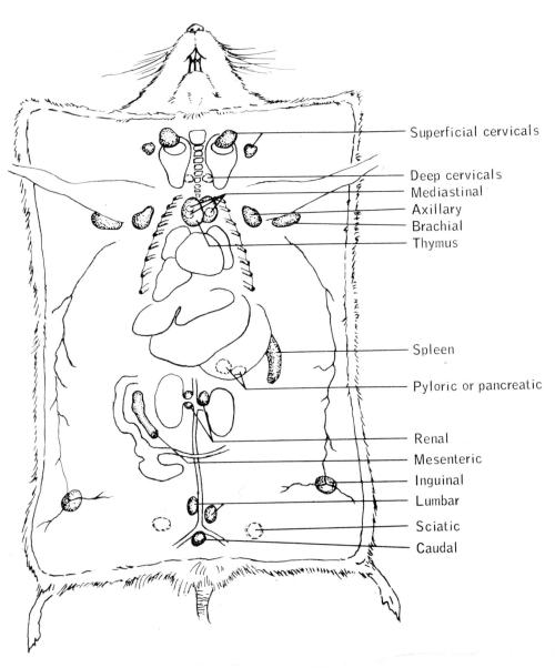
Diagram of Mouse Thymus, Spleen and Lymph Nodes The diagram below shows the locations of various lymphoid tissues in the mouse. Thymus and spleen are represented in red, while lymph nodes are color coded in green. The spleen sits in the peritoneal cavity on high under the rib cage on the left side. It is prominent and dark red in color.

Mouse anatomy diagram
Mouse Brain Diagram. whole brain low intensity pulsed ultrasound therapy whole brain low intensity pulsed ultrasound therapy markedly improves cognitive dysfunctions in mouse models of dementia crucial roles of endothelial nervous system explore the nerves with interactive the human nervous system - interact with diagrams and descriptions of the nervous system anatomy of the human body ... thicker and more obvious than the mouse's& d. Next sex your mouse. First locate the anus on the ventral side of the mouse near the base of the tale. Determine if testes (male gonads) are present. If yes, then your mouse is male; if no, then your mouse is most likely female.& & _____side& _____side& _____& Order your anatomy atlas from the AALAS Store!. Comparative Anatomy of the Mouse and Rat: a Color Atlas and Text provides detailed comparative anatomical information for those who work with mice and rats in animal research. Information is provided about the anatomical features and landmarks for conducting a physical examination, collecting biological samples, making injections of therapeutic ...
Mouse anatomy diagram. Amazon Com Ambesonne Human Anatomy Mouse Pad Diagram Of Human Hox Patterning Of The Vertebrate Rib Cage Development Bone Development In Laboratory Mammals Used In Developmental Animal Woodland Mouse Anatomy Animal Reference In 2019 Art The Anatomy Of The Laboratory Mouse Anatomy Of The Laboratory Mouse In Vivo Imaging Atlas On A High ... 14.4.2015 · The hand has several muscles. Some make broad, smooth movements, and others make small, finite movements. It’s the combination of the exterior and … When the screen pointer is over the icon of the object, the mouse button is clicked to grab it then you drag it to its new location. Drag and Drop a gesture in which the user selects a virtual object by Clicking it and dragging it to a different location or onto another virtual object. The Allen Mouse Brain Atlas includes a full-color, high-resolution anatomic reference atlas accompanied by a systematic, hierarchically organized taxonomy of mouse brain structures. In 2011, the reference atlas was updated to enable interactive online exploration of the atlas and to provide a deeper level of 3-D annotation for informatics ...
8.11.2019 · Hinge joints allow bones to move in one direction back and forth, much like the hinge on a door. This article looks at their anatomy and function and includes an interactive diagram. Start studying Mouse Anatomy. Learn vocabulary, terms, and more with flashcards, games, and other study tools. 10.10.2021 · Muscular System Anatomy Muscle Types. There are three types of muscle tissue: Visceral, cardiac, and skeletal. Visceral Muscle. Visceral muscle is found inside of organs like the stomach, intestines, and blood vessels. The weakest of all muscle tissues, visceral muscle makes organs contract to move substances through the organ. The Mouse Nervous System provides a comprehensive account of the central nervous system of the mouse. The book is aimed at molecular biologists who need a book that introduces them to the anatomy of the mouse brain and spinal cord, but also takes them into the relevant details of development and organization of the area they have chosen to study.
Cellular anatomy of the mouse primary motor cortex ... type-based wiring diagram of the mouse MOp-ul that will facilitate future analyses of motor control infrastructure across molecular, cellular ... Mouse liver lobes Source: Figures 2 and 3 in Harada, T., et al. "Liver and Gallbladder." Chapter 7 in Pathology of the Mouse. Edited by Robert Maronpot. Vienna, IL: Cache River Press, 1999. ISBN: 188989902X. Images removed for copyright reasons. This is the first interactive mouse necropsy site on the web. Designed to aid the researcher in learning and practicing the correct methods of retrieving tissues and examining mouse anatomy, this site is not only educational, but fun too!! Obviously, the procedures within this site can be useful in the necropsies of other species as well. The Mouse Limb Anatomy Atlas is a free, web-based, standardised reference of limb muscle, tendon and skeletal structures at embryonic day 14.5. The Atlas features interactive and annotated 2D and 3D models of the forelimb and hindlimb, showing over 60 individually segmented structures. This is the first complete reference tool for studying the ...
The Anatomy Of The Laboratory Mouse
Anatomy of a Neuron / Anatomy of a Synapse In part 1, students label a diagram of a neuron with structure and function (use Make a Mad, Mad, Mad Neuron as a source). In part 2, they describe what is happening in a labeled diagram of a synapse (use Crossing the Divide as a source).
The Anatomy Of The Laboratory Mouse
Figure 4: Key to mouse mammary tissue. Figure 5: Anatomy after Peritoneal Wall Incision 6. If the animal is male, the penis and the prepuital gland will lie on top of the muscle. By cutting between the prepuital gland and the muscle wall via the penis, the gland is left intact for inspection or preservation. 7.
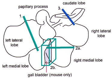
Revised Guides For Organ Sampling And Trimming In Rats And Mice Liver Gall Bladder Mouse Only
The pancreas of an adult mouse is surrounded by the stomach, the duodenum and proximal jejunum, the spleen and kidney, as shown in Fig. 8(B). The duodenum wraps around the head of the pancreas. Rodent pancreas is soft and diffuse compared with the human pancreas, and it is enclosed in the mesenteric adipose tissue completely. diagram
Mouse Anatomy
Mouse Anatomy Diagram. Share this worksheet. Can your preschooler name the parts of a mouse? Give her a dose of animal science with this fun cut-and-paste printable. Help her complete the mouse chart by showing her how to cut out the names of the parts and paste them in the right spaces. She'll get an introduction to animal anatomy and work on ...

Esophagus Function An Overview Sciencedirect Topics
Rodent skeletons come in all shapes and sizes, but they all have four extra-large front teeth powered by large, muscly jaws. The word "rodent" comes from the Latin word rodere, meaning "to gnaw," and these animals are experts at nibbling through tough plants to get at food and to find shelter.Their giant front teeth grow continuously throughout a rodent's life, because the constant ...

442 Mouse Anatomy Stock Photos Pictures Royalty Free Images Istock
Comparative Anatomy Chart This table contains a comparison between mouse and human anatomy. These pages were put together as a pilot demonstration by Dr. Robert Cardiff , UCD Center for Comparative Medicine with the collaboration of Dr. Michael Paulus, Oak Ridge National Laboratories, MicroCat Group , Dr. Allan Johnson, Duke University Center ...
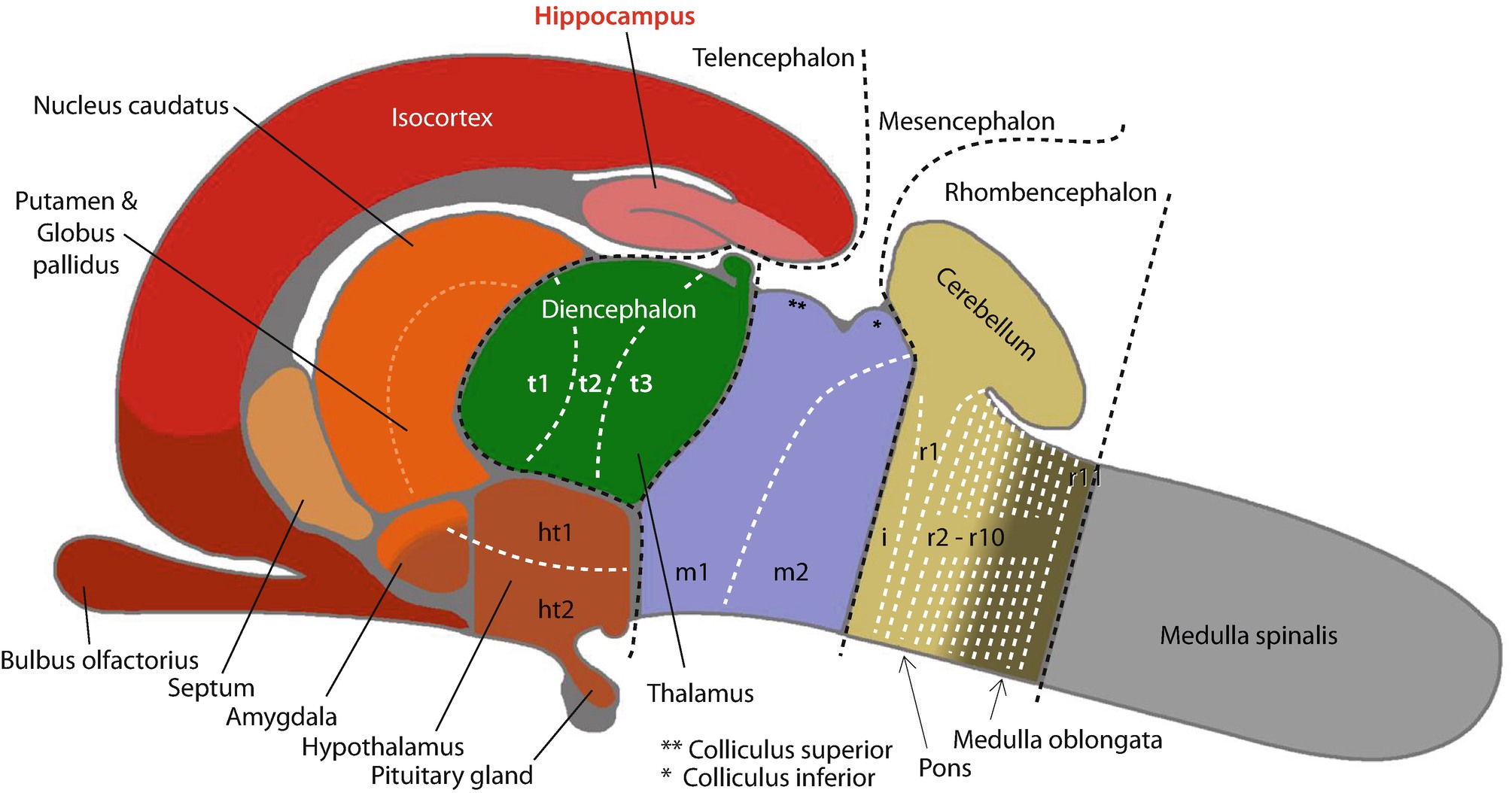
The Mouse Hippocampus Springerlink
The thicknesses and spheric and aspheric curvatures of the optic components were measured from cross-sections of frozen eyes of C57B1/6J mice. The equivalent refractive index of the crystalline lens was obtained from its back-vertex power in albumin. Refractive indices of the cornea, aqueous and vit …
2
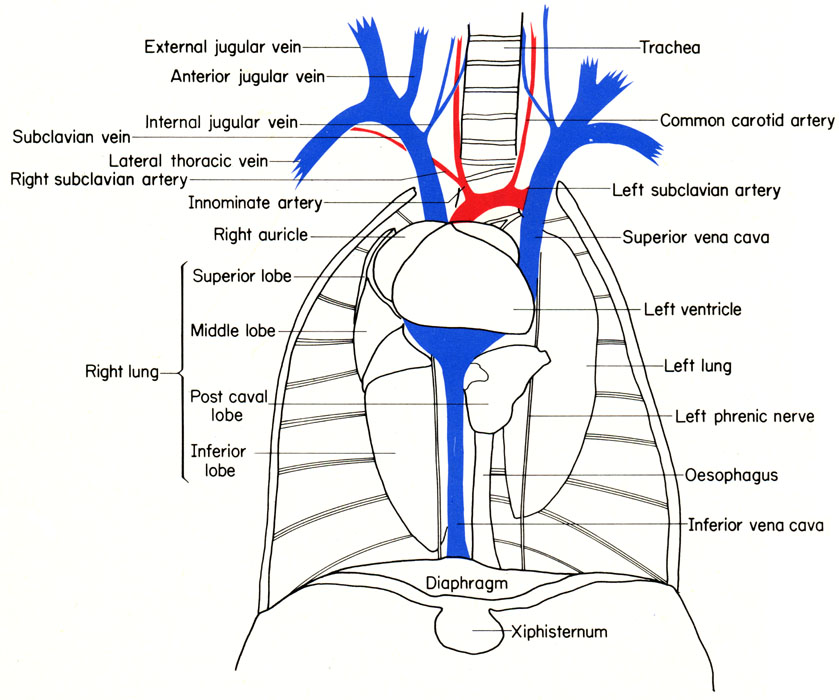
1 Oxygen-poor blood (shown in blue) flows from the body into the right atrium.. 2 Blood flows through the right atrium into the right ventricle.. 3 The right ventricle pumps the blood to the lungs, where the blood releases waste gases and picks up oxygen.. 4 The newly oxygen-rich blood (shown in red) returns to the heart and enters the left atrium.
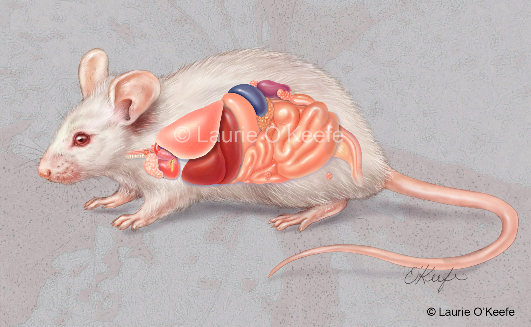
Rat Anatomy Archives Laurie O Keefe Illustration
Important points to remember for mouse brain histology : --Perfusion fixation is important to avoid artefacts, such as "dark neurons" --Do not leave in 70% alcohol for longer than 24 hours, to avoid vacuole artefact --both erythrocytes and degenerating neurons are autofluorescent. --To avoid hypostatic congestion in multiple tissues and to

Mouse Anatomy Stock Image Z918 0462 Science Photo Library
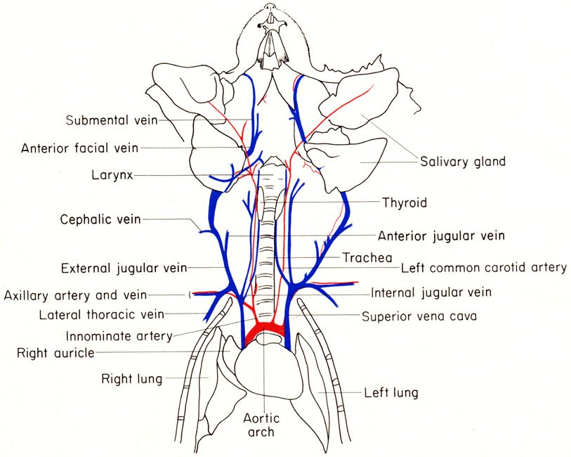
112. Blood supply of left kidney and suprarenal gland. 113. Male reproductive organs. 114. Blood supply of testis. 115. Male organs deflected to left to show blood supply.

Mouse Dissection Youtube
The nose is the body's primary organ of smell and also functions as part of the body's respiratory system. Air comes into the body through the nose. As it passes over the specialized cells of the ...
Mouse Printout Enchantedlearning Com
It should be noted that there are many more muscles in the body that are not addressed by this muscle anatomy diagram, however the muscles that are of primary interest from a fitness and exercise perspective are covered by this muscle anatomy diagram. Muscle Anatomy Diagram. Roll your mouse over any muscle in the diagram below to learn its name.

Jci The Placenta Transcriptional Epigenetic And Physiological Integration During Development
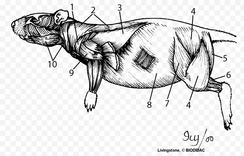
The Anatomy of the Laboratory Mouse Margaret J. Cook CONTENTS Contents: Abbreviated Title Page Foreword Introduction Externals 4. Mus musculus (LAC Grey strain). Adult male. 5. Vibrissae. 6. Left forepaw. 7. Right hind paw. 8. External genitalia. Female. 9. External genitalia. Male. Skeleton 10. Skeleton of LAC Grey mouse. 11. Dorsal aspect of ...

Brainsitu Gallery Cortex
Research & reviews for your most important home health purchases. Turn to our experts for everything you need to know about at-home testing and telemedicine.
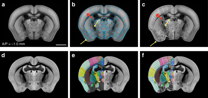
Enhanced And Unified Anatomical Labeling For A Common Mouse Brain Atlas Nature Communications
Labeled cross-sectional anatomy of the mouse on micro-CT. These images of a normal female Swiss mouse have been acquired with a laboratory-based microCT system (nanoScan PET/CT Mediso (Budapest, Hungary) with an operation voltage of 50kVp and a 0,14mm pitch, with an intravenous injection of 2ml of Visipaque (320mg d'I/ml), at CERIMED (Centre Européen de Recherche en Imagerie Médicale ...

File Basic Mouse Liver Anatomy Svg Wikimedia Commons
5.11.2019 · Female anatomy includes the external genitals, or the vulva, and the internal reproductive organs. This article looks at female body parts and their functions, and it provides an interactive diagram.

The Anatomy Of The Laboratory Mouse
Anatomy (Greek anatomē, 'dissection') is the branch of biology concerned with the study of the structure of organisms and their parts. Anatomy is a branch of natural science which deals with the structural organization of living things. It is an old science, having its beginnings in prehistoric times. Anatomy is inherently tied to developmental biology, embryology, comparative anatomy ...

Human And Mouse Brain Comparison Metal Print By Evan Oto
The Anatomy Of The Laboratory Mouse The anatomy of the laboratory mouse. Mouse dissection diagram. Diagram for location of incisions. The anatomy of the laboratory mouse margaret j. Thymus and spleen are represented in red while lymph nodes are color coded in green.

Amazon Com Ambesonne Science Mouse Pad Structure Of The Heart Human Body Anatomy Organ Veins Cardiology Rectangle Non Slip Rubber Mousepad Standard Size Coral Red Office Products
The Laboratory Mouse, Second Edition is a comprehensive book written by international experts. With inclusions of the newly revised European standards on laboratory animals, this will be the most current, global authority on the care of mice in laboratory research.
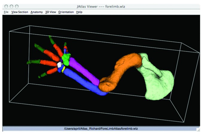
The Mouse Limb Anatomy Atlas An Interactive 3d Tool For Studying Embryonic Limb Patterning Bmc Developmental Biology Full Text
Order your anatomy atlas from the AALAS Store!. Comparative Anatomy of the Mouse and Rat: a Color Atlas and Text provides detailed comparative anatomical information for those who work with mice and rats in animal research. Information is provided about the anatomical features and landmarks for conducting a physical examination, collecting biological samples, making injections of therapeutic ...

Amazon Com Adowyee Gaming Mouse Pad Tooth Of Diagram For Anatomy Human Mouth Open Dental 9 5 X7 9 Nonslip Rubber Backing Computer Mousepad For Notebooks Mouse Mats Office Products
thicker and more obvious than the mouse's& d. Next sex your mouse. First locate the anus on the ventral side of the mouse near the base of the tale. Determine if testes (male gonads) are present. If yes, then your mouse is male; if no, then your mouse is most likely female.& & _____side& _____side& _____&
2
Mouse Brain Diagram. whole brain low intensity pulsed ultrasound therapy whole brain low intensity pulsed ultrasound therapy markedly improves cognitive dysfunctions in mouse models of dementia crucial roles of endothelial nervous system explore the nerves with interactive the human nervous system - interact with diagrams and descriptions of the nervous system anatomy of the human body ...

Cytoarchitecture Of The Spinal Cord Of The Postnatal P4 Mouse Sengul 2012 The Anatomical Record Wiley Online Library
Anatomy Of The Human And The Mouse Pancreas A The Parts Of The Human Download Scientific Diagram

Development Of The Kidney In Mouse And Human Gudmap

Human And Mouse Brain Comparison Photograph By Evan Oto
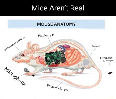
Mice Aren T Real Mice Aren T Real Mouse Anatomy Raspberry Pi D O
1

Investigation Rat Dissection Biology Libretexts

Mouse Anatomy

Mouse Bat Unit Study Mega Printable Bundle Posters Etsy Bats Unit Study Bats Activities Bats Unit
Mouse Anatomy Facebase
2

2 A And B External Mouse Anatomy Download Scientific Diagram

Hand Drawn Illustration Of Human Heart Anatomy Educational Diagram Showing Blood Flow With Main Parts Labeled Vector Illustration Easy To Edit Mouse Pad Id 218235622

Mouse Brain Anatomy Dorsal And Ventral Views

Mouse Brain Anatomy Sagittal View
1

Mouse Anatomy

Mouse Diagram Quizlet
Heart Rat Dissection Muscle Anatomy Human Body Cartoon Mouse Free Png
2
Comments
Post a Comment