42 muscle fiber diagram labeled
Internal Anatomy of Skeletal Muscle Fibers - GetBodySmart Skeletal Muscle Fiber Location and Arrangement. are located inside muscles, where they are organized into bundles called […] General Anatomy of Skeletal Muscle Fibers. An interactive quiz about the general anatomy of skeletal muscle fibers, featuring illustrations-based multiple choice questions. Labeled Sarcomere Diagram The thin filaments Look at the diagram above and realize what happens as a muscle contracts. As will soon be described, the functional unit of a skeletal muscle fiber is the sarcomere, a highly organized arrangement of the contractile myofilaments actin .Play this quiz called Label the Sarcomere and show off your skills.
Sarcomere Diagram Labeled - schematron.org Skeletal muscles are composed of tubular muscle cells which. Each myofibril is made up of contractile sarcomeres AND Drawing labelled diagrams of the structure of a sarcomere.Summary. A little over 50 years ago, Sydney Brenner had the foresight to develop the nematode (round worm) Caenorhabditis elegans as a genetic model for understanding questions of developmental biology and neurobiology.

Muscle fiber diagram labeled
open.oregonstate.education › aandp › chapter19.2 Cardiac Muscle and Electrical Activity – Anatomy ... Figure 19.2.1 – Cardiac Muscle: (a) Cardiac muscle cells have myofibrils composed of myofilaments arranged in sarcomeres, T tubules to transmit the impulse from the sarcolemma to the interior of the cell, numerous mitochondria for energy, and intercalated discs that are found at the junction of different cardiac muscle cells. Muscles - Cabarrus County Schools 1. There are over 1,000 muscles in your body. 2. Skeletal, or voluntary, muscles are the muscles you can control. 3. Ligaments connect muscles to bones. 4. Your heart is a muscle. 5. A muscle gets strained when it is stretched too much. 6. A sprain happens when a tendon is stretched too much. 7. Muscles that are not used can get smaller and weaker 8. Muscle Fiber Labeling Quiz - PurposeGames.com This is an online quiz called Muscle Fiber Labeling There is a printable worksheet available for download here so you can take the quiz with pen and paper. Your Skills & Rank
Muscle fiber diagram labeled. Sarcomere Diagram Labeled As will soon be described, the functional unit of a skeletal muscle fiber is the sarcomere, a highly organized arrangement of the contractile myofilaments actin . Draw your own diagram of two sarcomeres. The first should be of a relaxed muscle. The second should be of a contracted muscle. Label the Z line, M line. Muscle fibres - Energy systems in muscle cells - Higher ... Muscle fibres. The human body contains different types of muscle fibres: Cardiac muscle - unique to the heart - never tires. Involuntary muscles - work our internal organs - outside our control ... Muscle Fiber Structure Inner Parts Anatomical Stock Vector ... Download for free. Royalty-free stock vector ID: 2067131960. Muscle fiber structure and inner parts anatomical description outline diagram. Labeled educational medical organ scheme with myofibril, sarcolemma, sarcoplasmic reticulum location vector illustration. V. PDF Muscular System Tour Skeletal Muscle Muscle Fiber (muscle cell) Myofibril Contracted. Myofibril relaxed. Muscle Cell. Fascicle (group of muscle cells) A. J. G. ... The Muscles … a back view. H. F. Use your front view and back view diagrams to label these muscles Gluteus medius Label: place the letter next to the name. Gluteus maximus . Latissimus dorsi Pectoralis major ...
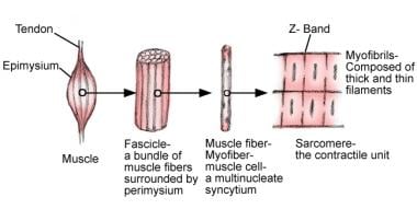
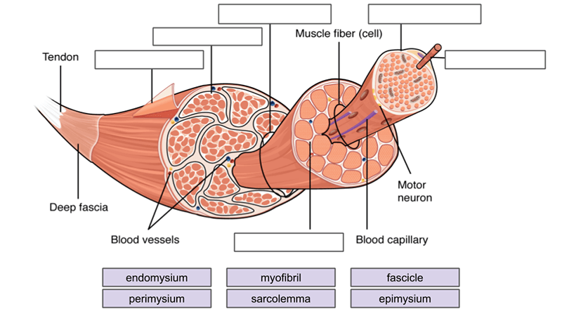
PDF THE MUSCULAR SYSTEM - University of Cincinnati Muscle fibers are organized into bundles supplied by blood vessels and innervated by motor neurons. Muscle structure. Skeletal (striated or voluntary) muscle consists of densely packed groups of hugely elongated cells known as myofibers. These are grouped into bundles (fascicles). General Anatomy of Skeletal Muscle Fibers - GetBodySmart Skeletal Muscle Fiber Location and Arrangement are located inside muscles, where they are organized into bundles called […] Internal Anatomy of Skeletal Muscle Fibers en.wikipedia.org › wiki › White_ramus_communicansWhite ramus communicans - Wikipedia Structure. The white rami communicantes are the preganglionic sympathetic outflow from the spinal cord. The cell bodies for the preganglionic sympathetic myelinated fibers in the white rami communicantes lie in the ipsilateral (same sided) intermediolateral cell column in the spinal cord which extends from T1-L2. › cell › cell-structureCell: Structure and Functions (With Diagram) Another kind of fiber found in cytoplasm of most eukaryotes. Involved in muscle contraction, cell support, pinching off of daughter cells after mitosis. Extracellular matrix (ECM): Animal cells do not have cell walls, but have ECM, i.e., a meshwork of macromolecules outside plasma membrane.
sciencetrends.com › labeled-neuron-diagramLabeled Neuron Diagram - Science Trends May 29, 2019 · Pseudounipolar: Single axon/dendrite fiber with a soma protrusion Dividing and classifying the different kinds of brain cells is a massively complex task. There is currently no consensus about how many kinds of neurons exist in the brain , but scientists have identified 3 major kinds of neurons in the spinal cord: sensory, motor, and interneurons. Label structure of skeletal muscle Diagram | Quizlet Label structure of skeletal muscle Diagram | Quizlet. Muscle Cell Labelled Diagram : Muscles Labeling : - Pic ... (a) on the diagram, use words from the box to name the structures labelled . One muscle cell is known as a muscle fibre, and its cell surface membrane is known as the sarcolemma. A pie slice diagram shows the proportion of water to typical chemical . Hence giving these nerve and muscle cells have the ability to move. Fresh Blank Muscular System Diagram - Labelco The free muscular system labeling sheet includes a blank diagram to label some of the main muscles in the body. Human Anatomy And Physiology. Anatomy of the Eye Provide the labels for the indicated parts on the diagram of an eye. One is in color and the other is in black and white. Numbering Worksheets for Kids.
Muscle Contraction Diagram (labelled) - Stock Image - C043 ...
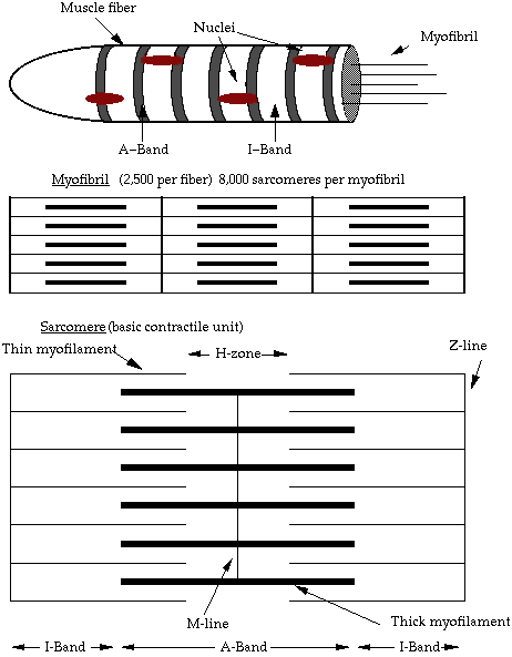
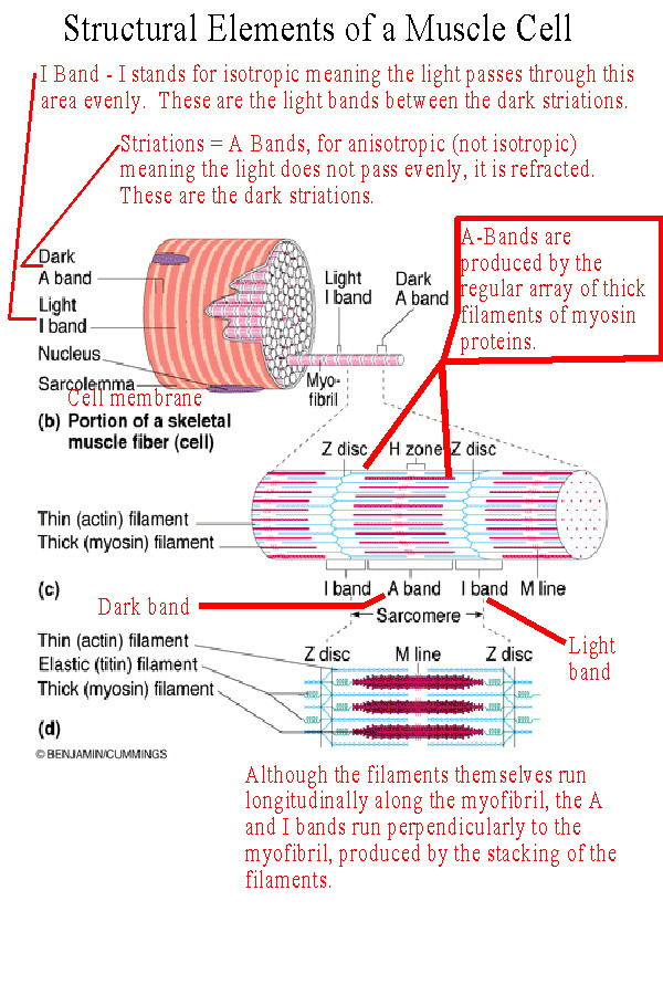
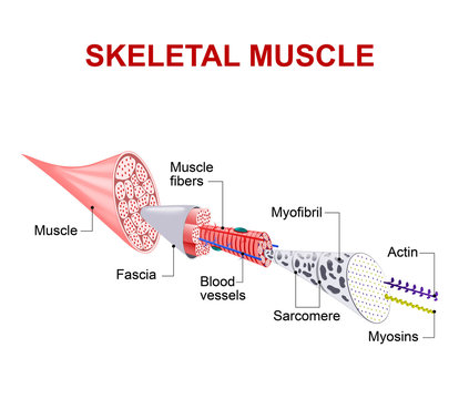
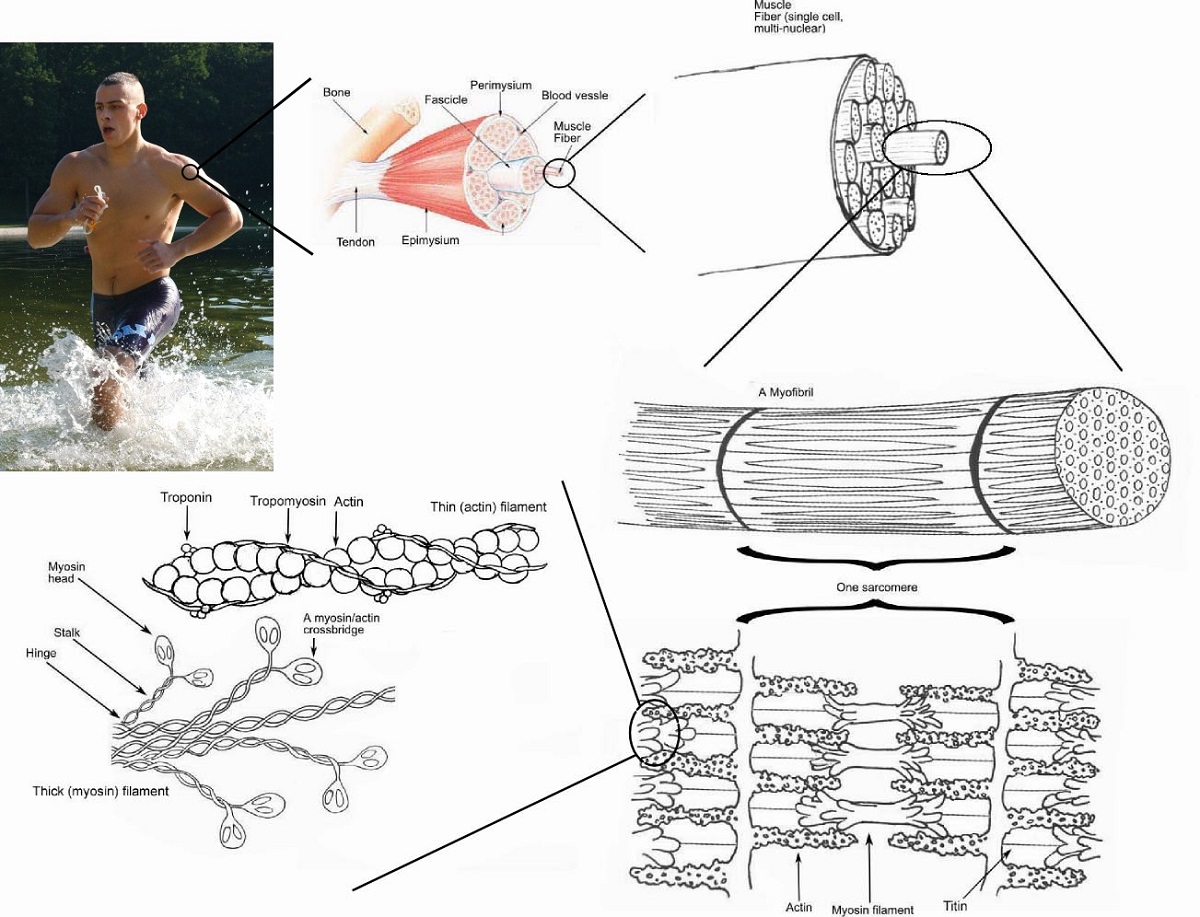
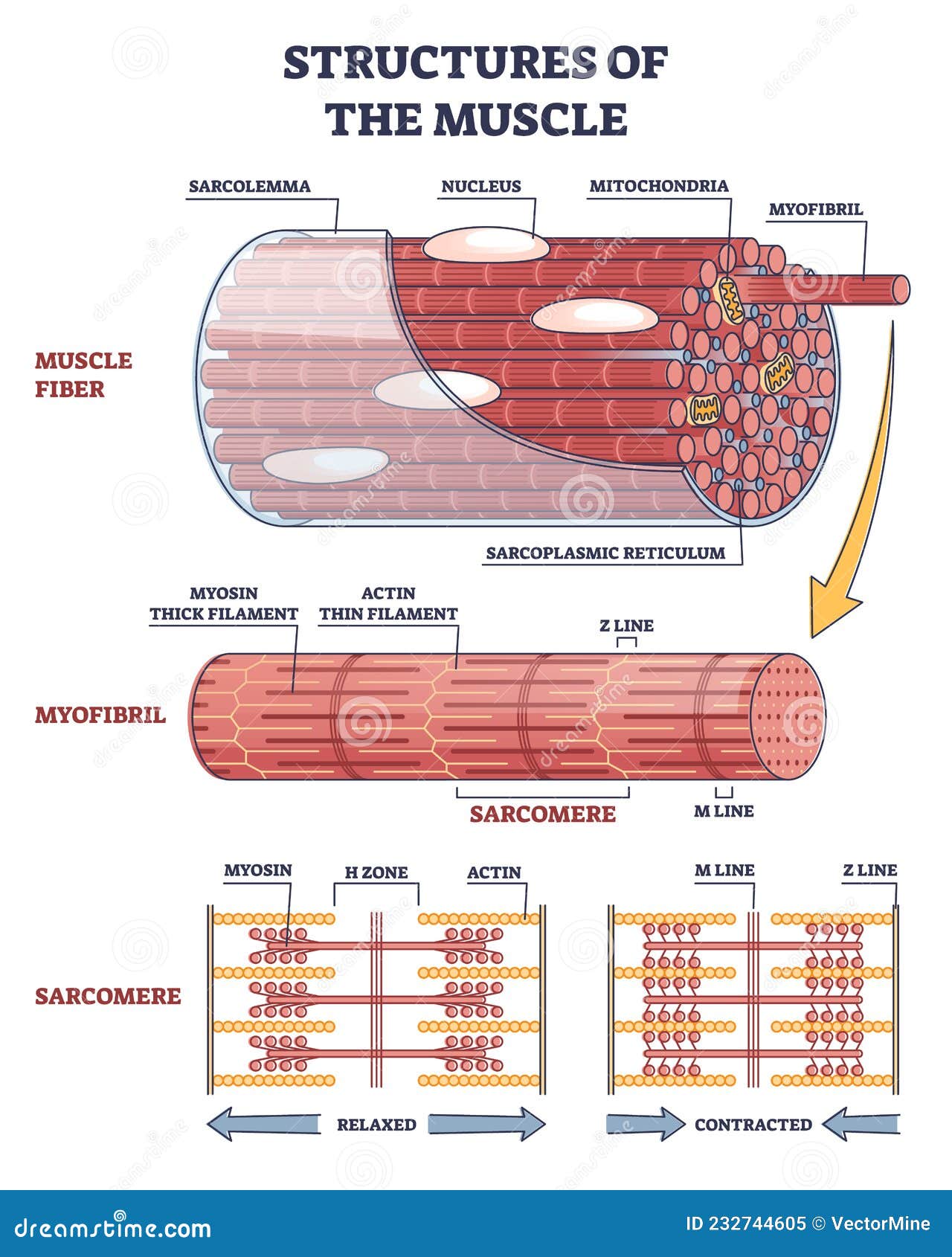
Muscle Fiber Anatomy In Detail Diagram of part of a muscle fiber showing the myofibrils. One myofibril extends from the cut end of the fiber. Small part of one myofibril enlarged to show the myofilaments responsible for the banding pattern. Each sarcomere extends from one Z disc to the next. Enlargement of one sarcomere (sectioned lengthwise).
Structure and Composition of Muscle - Meat Science
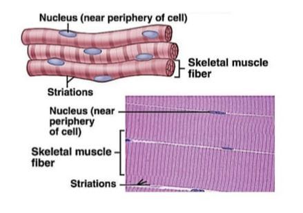
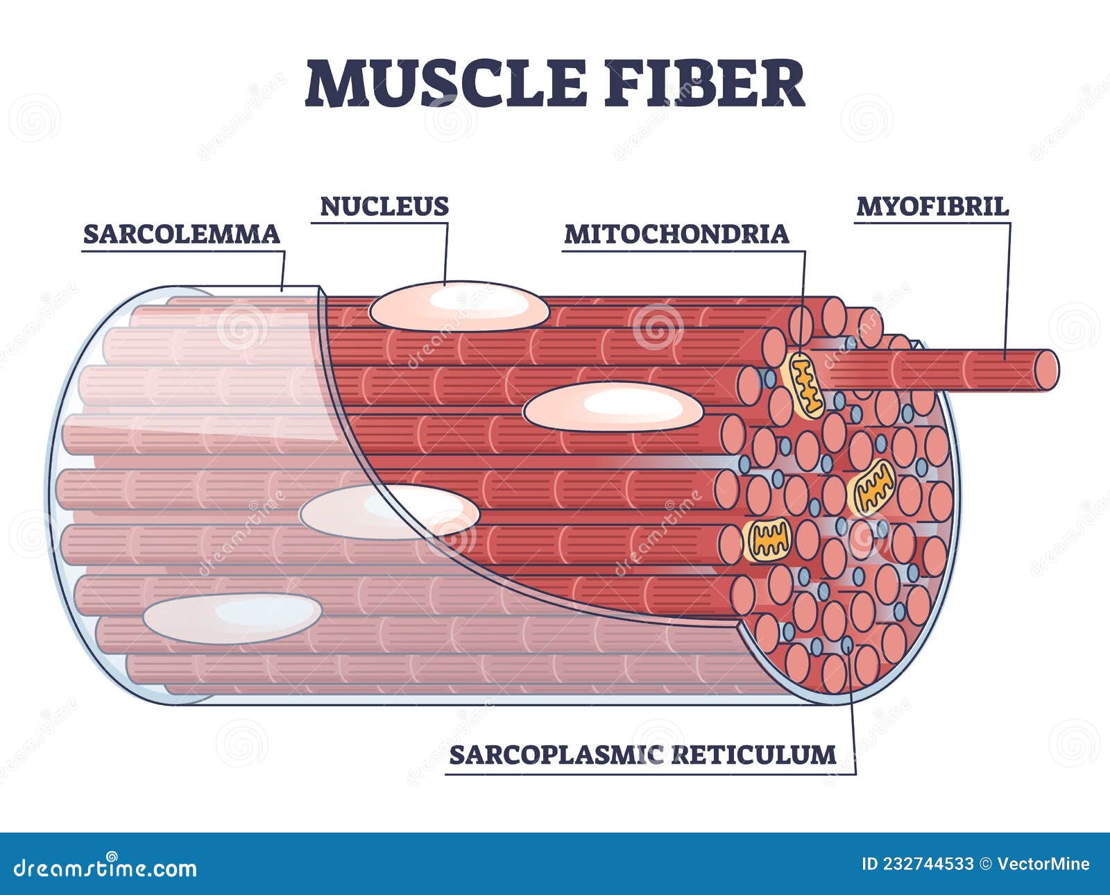
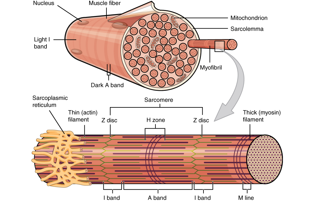
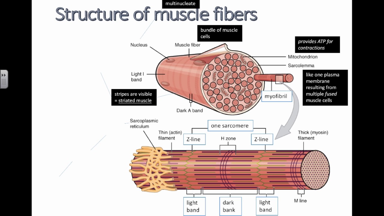
Skeletal Muscle Fiber Structure and Function | Open ... Each skeletal muscle fiber is a skeletal muscle cell. Within each muscle fiber are myofibrils, long cylindrical structures that lie parallel to the muscle fiber. Myofibrils run the entire length of the muscle fiber. They attach to the plasma membrane, called the sarcolemma, at their ends, so that as myofibrils shorten, the entire muscle cell contracts (Figure 16.18).

Muscle Fiber Anatomy Quiz
› pmc › articlesStructure and Function of the Skeletal Muscle Extracellular ... Sep 01, 2011 · The Muscle Endomysium . An exception to the casual sampling performed in most ECM studies is the systematic and quantitative description of the muscle endomysial ECM reported for feline and bovine muscle by Purslow and Trotter. 2, 3 They showed that a highly ordered network surrounds individual muscle fibers that deforms nonlinearly with increasing sarcomere length.
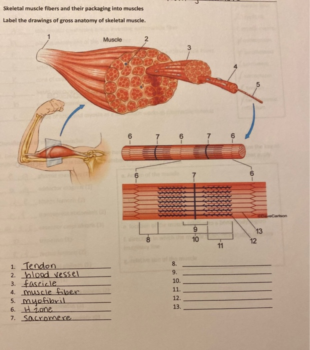
Solved Skeletal muscle fibers and their packaging into ...
Skeletal Muscle Histology Slide Identification and Labeled ... Skeletal muscle histology description with labeled diagram #1. Epimysium of muscle fibers - a dense, irregular connective tissue layer surroundings the entire skeletal muscle... #2. Perimysium of muscle fibers - you know, a muscle fascicle is a small bundle of muscle fibers. A dense, irregular... ...
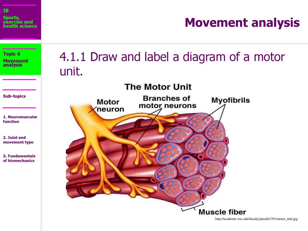
4.1.1 Draw and label a diagram of a motor unit. - ppt download
skeletal muscle fiber labeled Diagram | Quizlet skeletal muscle fiber labeled Diagram | Quizlet. Start studying skeletal muscle fiber labeled. Learn vocabulary, terms, and more with flashcards, games, and other study tools. Search.
Muscle Tissue | Basicmedical Key
Muscle Charts of the Human Body - PT Direct For your reference value these charts show the major superficial and deep muscles of the human body. Superficial and deep anterior muscles of upper body Superficial and deep posterior muscles of upper body
Topic 11.2: Movement - AMAZING WORLD OF SCIENCE WITH MR. GREEN
bodytomy.com › nodes-of-ranvier-location-functionNodes of Ranvier: Location And Function (With a Labeled Diagram) Neurons are the most fundamental units of the nervous system. They connect the brain and spinal cord to every organ and muscle fiber in the body. Neurons constitute our body’s response mechanism; they receive sensory stimulation from the sense organs, and execute an appropriate response by producing the required muscular movement.

Smooth Muscle | Anatomy and Physiology I
Skeletal muscle fiber - human anatomy organs SKELETAL MUSCLE FIBER STRUCTURE A 12 inch muscle fiber is not uncommon in the human body, which another specific unique characteristic of skeletal muscles. The diameter of a single muscle fiber can range anywhere from 10 micrometers to 100 micrometers. Sarcolemma, a unique cell membrane, completely encases each individual muscle fiber.

10.2 Skeletal Muscle – Anatomy & Physiology
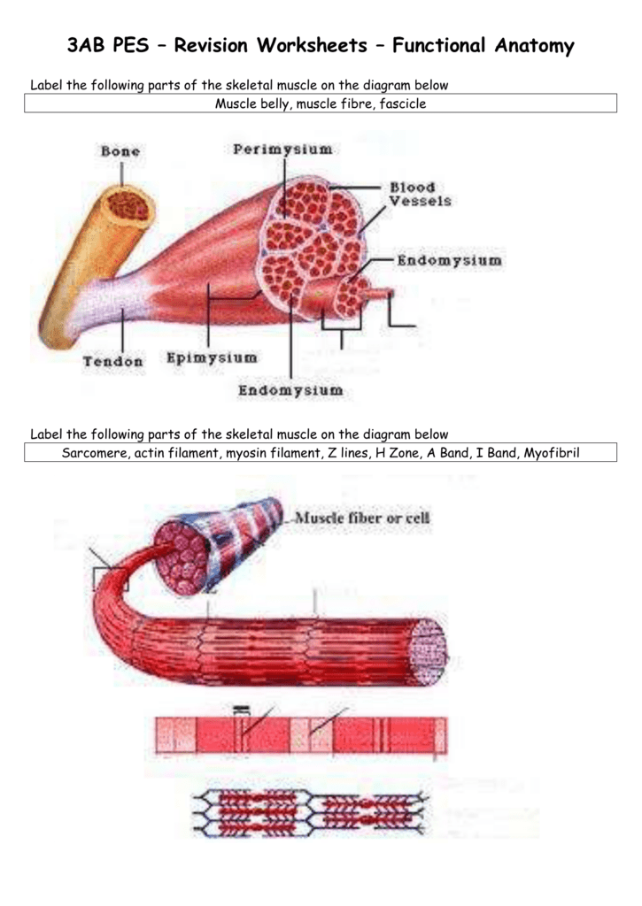
PDF Muscular System Worksheet - Anatomy Academy Label the anatomy of a muscle fiber. Muscle fiber (cell) vessel 12. Label 1-7 on the following diagram. Use the word bank below. Striated (Skeletal) Muscle 8. Word bank: Sarcomere, Myosin, Z line, A band, I band, H zone, Actin. ANAT 113 Fill in the blanks: filament attached to Z line.
BIOL 237 Class Notes - Muscle Cells & Muscle Physiology
PDF Draw and label a diagram of a motor unit. Neuromuscular ... How do we determine our muscle fiber type? 1. Muscle biopsy (best method) 2. Testing an athlete's muscle groups for different muscle fiber properties. Example: establish an RM (repetition maximum) of any exercise. lift 80% of 1RM as many times as possible. 7 or less reps most likely more than 50%FT fibers 12 or more reps most likely more than ...

Skeletal Muscle Fiber label part 1 Diagram | Quizlet
› tissues-2 › muscularProperties of the Cardiac Muscle | Cardiovascular System ... Excitation-Contraction Coupling in Cardiac Muscle: Is the mechanism by which action potential causes myofibrils of cardiac muscle to contract. When action potential passes over cardiac muscle membrane, it also spreads to interior of cardiac muscle fiber along membranes of transverse (T) tubules.
ATP muscle contraction cycle vector illustration labeled ...
Diy Skeletal Muscle Diagram Worksheet - Labelco Label Muscles Worksheet Muscle Diagram Muscular. Skeletal or voluntary muscles are the muscles you can control. Complete this mind map about muscles bones joints or other. These free human body worksheets are perfect for kindergartners grade 1 grade 2 grade 3 grade 4 grade 5 and grade 6 students. Using Figure 61 match the following.
Muscle Fiber Structure and Inner Parts Anatomical Description ...
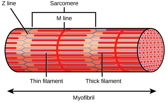
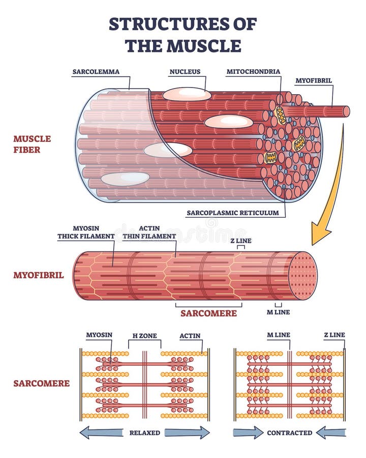
10.2 Skeletal Muscle - Anatomy & Physiology The plasma membrane of muscle fibers is called the sarcolemma (from the Greek sarco, which means "flesh") and the cytoplasm is referred to as sarcoplasm (Figure 10.2.2). Within a muscle fiber, proteins are organized into structures called myofibrils that run the length of the cell and contain sarcomeres connected in series. Because myofibrils are only approximately 1.2 μm in diameter, hundreds to thousands (each with thousands of sarcomeres) can be found inside one muscle fiber.
Gross and Microscopic Anatomy of Skeletal Muscle
Muscle Fiber Anatomy Quiz - PurposeGames.com This is an online quiz called Muscle Fiber Anatomy. There is a printable worksheet available for download here so you can take the quiz with pen and paper.

muscle fiber diagram | MUSCULAR SYSTEM with blanks | Muscle ...
20 Unlabeled Muscle Diagram Worksheet | Worksheet From Home 20 Unlabeled Muscle Diagram Worksheet. Label Muscles Worksheet unlabeled male reproductive system, unlabeled muscle fiber, unlabeled muscles, unlabeled muscular system image, unlabeled male reproductive system diagram, via: pinterest.com. Numbering Worksheets for Kids. Kids are usually introduced to this topic matter during their math education.

draw a well labelled diagram to show the difference in three ...
Muscle Fibers: Anatomy, Function, and More Muscle fibers are single muscle cells. When grouped together, they work to generate movement of your body and internal organs. You have three types of muscle tissue: skeletal, smooth, and cardiac.

Gross Anatomy Of Skeletal Muscle The Muscular System Micro ...
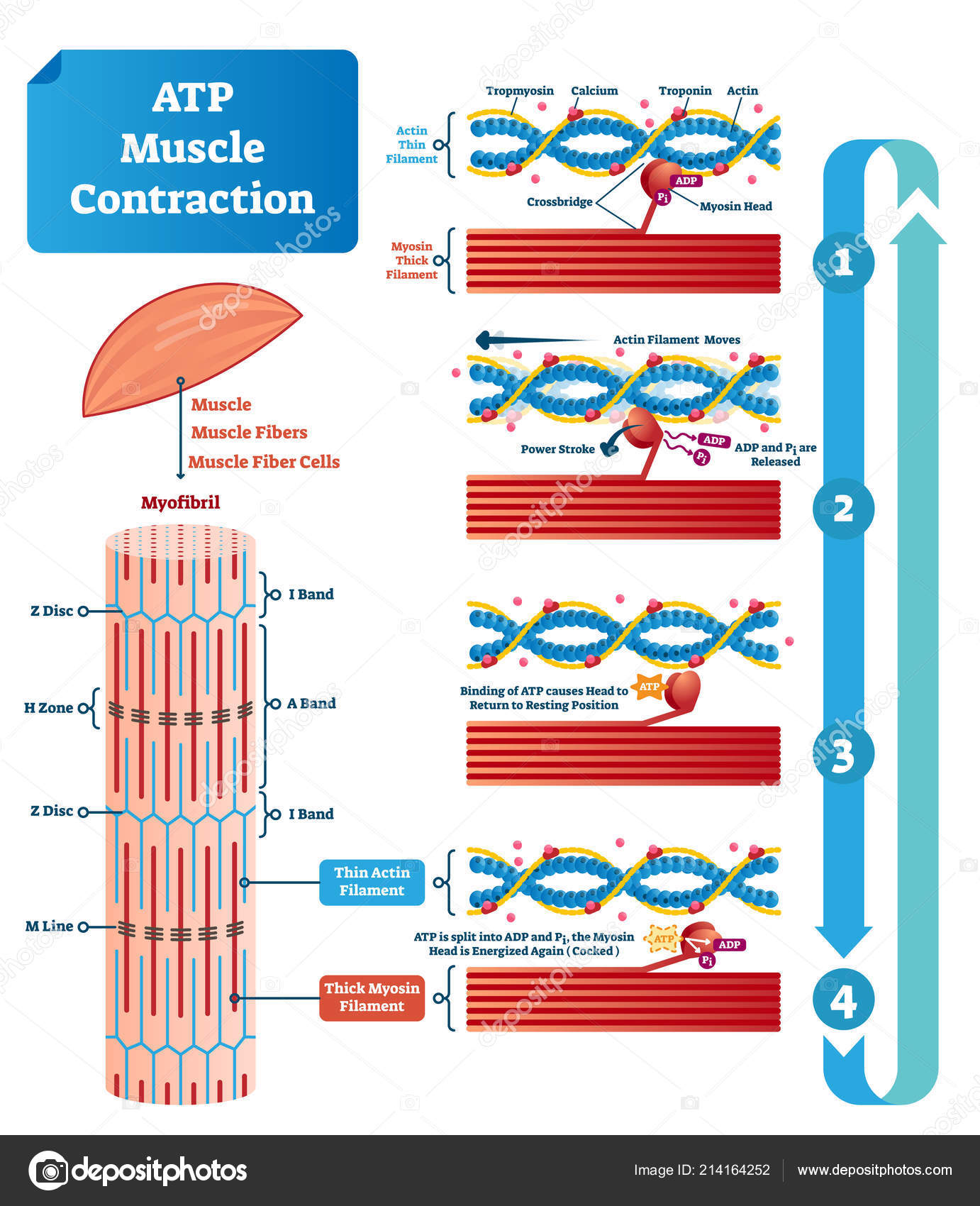
10.3 Muscle Fiber Excitation, Contraction, and Relaxation ... Diagram the process of cross-bridge cycling; The Neuromuscular Junction. The process of muscle contraction begins at the site where a motor neuron's terminal meets the muscle fiber—called the neuromuscular junction (NMJ). Every skeletal muscle fiber in every skeletal muscle is innervated by a motor neuron at a NMJ. Excitation signals from ...
Skeletal Muscle - Structure and Histology: Overview, Myofiber ...
PDF Anatomy Review: Skeletal Muscle Tissue • Are continuous with the tendons at the ends of the muscle. • Label this diagram: Page 7. Internal Structure of a Skeletal Muscle Cell • Label this diagram: Muscle fibers: Alternative name for skeletal muscle cells. • Nucleus: Contains the genetic material. • Sarcolemma: Plasma membrane of the muscle cell.
Structure of Muscle Fibers (IB Biology)
Muscle Fiber Labeling Quiz - PurposeGames.com This is an online quiz called Muscle Fiber Labeling There is a printable worksheet available for download here so you can take the quiz with pen and paper. Your Skills & Rank
Skeletal muscle - Structure - Contraction - TeachMePhysiology
Muscles - Cabarrus County Schools 1. There are over 1,000 muscles in your body. 2. Skeletal, or voluntary, muscles are the muscles you can control. 3. Ligaments connect muscles to bones. 4. Your heart is a muscle. 5. A muscle gets strained when it is stretched too much. 6. A sprain happens when a tendon is stretched too much. 7. Muscles that are not used can get smaller and weaker 8.

Human Physiology - Muscle
open.oregonstate.education › aandp › chapter19.2 Cardiac Muscle and Electrical Activity – Anatomy ... Figure 19.2.1 – Cardiac Muscle: (a) Cardiac muscle cells have myofibrils composed of myofilaments arranged in sarcomeres, T tubules to transmit the impulse from the sarcolemma to the interior of the cell, numerous mitochondria for energy, and intercalated discs that are found at the junction of different cardiac muscle cells.
Muscle Fibre Images – Browse 7,793 Stock Photos, Vectors, and ...

Muscle fascicle - Wikipedia
Sarcolemma Stock Photos, Stock Images and Vectors | Stockfresh
Muscular System- Part 1

Muscle Structure And Control Of Contraction - Muscle System ...

B1 Muscles and Movement | Brent Cornell

1 Composition of skeletal muscle tissue. (a) Whole muscle ...
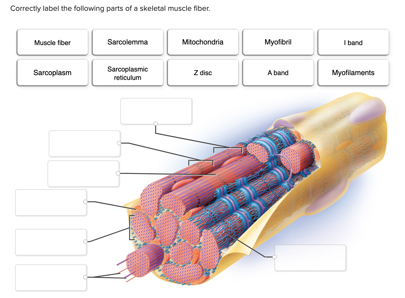
Solved Correctly label the following parts of a skeletal ...
Skeletal Muscle: Definition, Function, Structure, Location ...

Muscle Strains and Tears

Skeletal muscle fiber Diagram | Quizlet
Structures of Muscle with Fiber, Myofibril and Sarcomere ...

Muscle Fiber Labeling Diagram | Quizlet
Muscles Labeling
Muscles Labeling

STRUCTURE OF A SKELETAL MUSCLE FIBER Diagram | Quizlet
3AB PES – Revision Worksheets – Functional - PE Studies

The Structure of a skeletal Muscle fiber | Biology lessons ...

Muscle Fiber Contraction and Relaxation | Anatomy and ...
Structures of Muscle with Fiber, Myofibril and Sarcomere ...

Neuromuscular junction vector illustration scheme. Labeled cell infographic art print poster


Comments
Post a Comment