43 cell membrane diagram labeled
Antibody- Structure, Classes and Functions. Antibody (Ab) also know as Immunoglobulin (Ig) is the large Y shaped protein produced by the body's immune system when it detects harmful substances, called antigens like bacteria and viruses. The production of antibodies is a major function of the immune system and is carried out by a type of white ... Metabolism and energetics, structure and function of biomolecules, cell structure and function, animal development. Second course in a three-quarter series (BIOL 180, BIOL 200, BIOL 220). Prerequisite: minimum grade of 1.7 in either BIOL 180, B BIO 180, T BIOL 120, or TESC 120; either CHEM 143, CHEM 145, CHEM 223, CHEM 237, or OCEAN 295, or ...
Fig. 2. DNAgel synthesis and processability. ( A) Schematic of the synthesis process. DNAgel precursor is prepared by dissolving dehydrated DNA strands and then chemically crosslinked by poly (ethylene glycol) diglycidyl ether (PEGDE), forming a 3D network at room temperature. ( B) Scanning electron microscope image of freeze-dried DNAgel.

Cell membrane diagram labeled
The respiratory system of the pig commences at the nostrils which lead into two nasal passages. These contain the dorsal and ventral turbinate bones. (Fig.1-8). The ventral turbinates consist of four thin main bones, two on each side separated by a cartilaginous septum. You can imagine these as four hair curlers placed inside the nose. (BOSTON) — The inherited progressive disorder cystic fibrosis (CF) causes severe damage to the lungs, and other tissues in the body by affecting the cells that produce mucus, sweat, and ... Lesson Summary. Keratin is an important protein in the epidermis. Keratin has two main functions: to adhere cells to each other and to form a protective layer on the outside of the skin. In ...
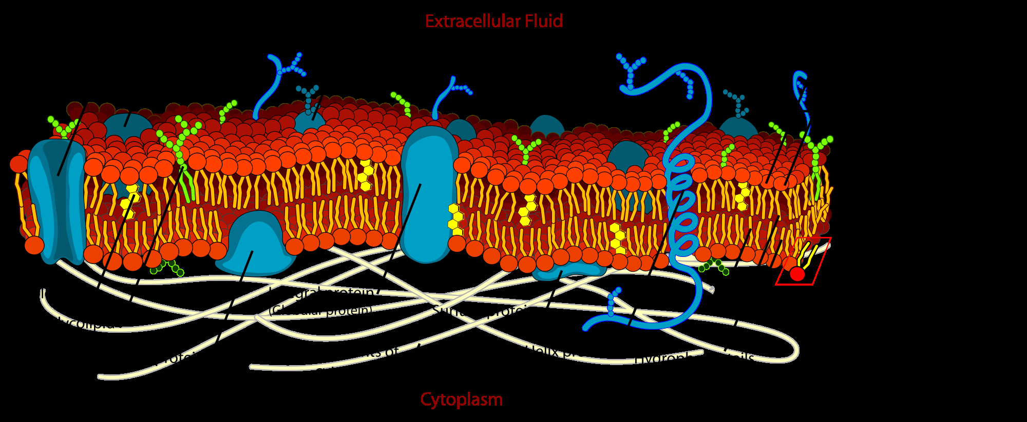
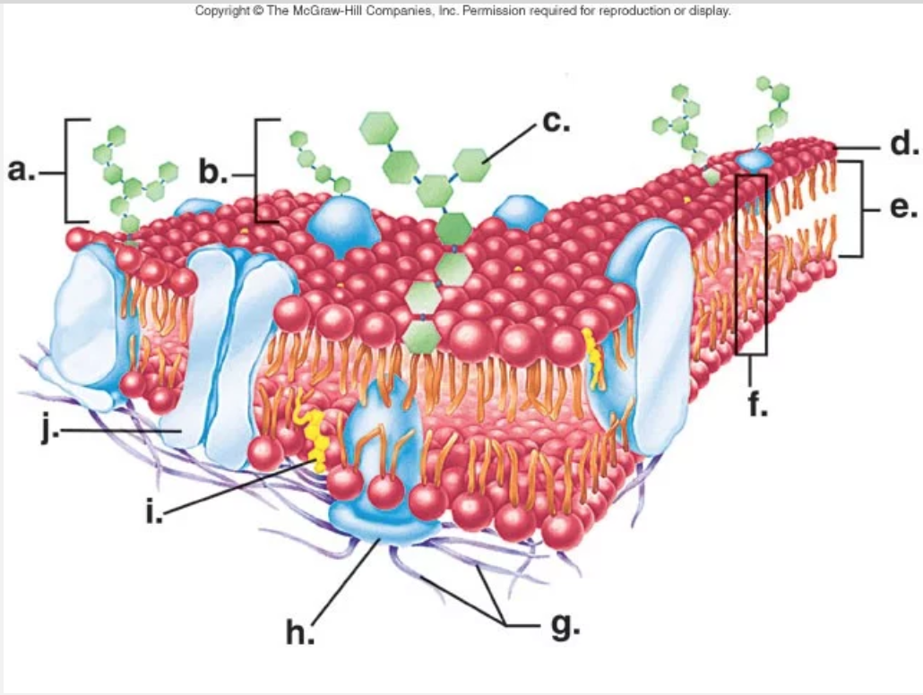
Cell membrane diagram labeled. New research reveals that specialized cells within neural circuitry that triggers complex learning in songbirds bears a striking resemblance to a type of neural cell associated with the ... Students who have that structure in their cell must fill in the blank on their bingo sheet. The first student to fill in all the blanks on his/her cell raises his/her hand. To be the winner, the student must have labeled each structure properly and be able to tell the class the function of each cell structure and what type of cell it is. In other words, a diagram of the membrane (like the one below) is just a snapshot of a dynamic process in which phospholipids and proteins are continually ...The cell membrane review · Fluid mosaic model of cell... · Practice The chick pierces the inner shell membrane and breathes in the air cell. Gas exchanges occur through the shell, which is porous. The chick is ready to hatch. Piercing of the shell begins. Day 21: The chick uses its wing as a guide and its legs to turn around and pierce the shell in a circular way by means of its egg-tooth.
Cell Membrane: Structure and Function Quiz. Cell Membrane: Structure and Function. Quiz. 1. The cell membrane is the outermost part of an animal cell, and, is situated underneath the cell wall in plant cells. What is another name for the cell membrane? Liquid Membrane. Conjoint Membrane. Pastoral Membrane. A squamous cell bladder carcinoma with karyotypic abnormalities reminiscent of transitional cell carcinoma. J Urol. 1989 Aug. 142 (2 pt 1):374-6. . Vanni R, Scarpa RM, Nieddu M, Usai E ... Components of Plasma Membranes. The plasma membrane protects the cell from its external environment, mediates cellular transport, and transmits cellular signals ... Study digs up roles bacteria play in global carbon cycle. Bacteria convert plant biomass, such as this corn organic matter, into soil, which adds to soil fertility and stores carbon in the ground ...
The Blood-Testis Barrier . The blood-brain barrier is a basic anatomical concept, with implications for disease and treatment. This system protects the brain and makes it difficult for many toxins, drugs, and other substances to affect it. There is a similar barrier in the testes that is referred to as the blood-testis barrier. Magnified view of the cyan boxed area marked. The orange and red asterisks indicate RSV glycoprotein fluorescent signals. Magnified cryo-EM montage view of Nanogold-Alexa488 immuno-labeled RSV particles. The RSV filaments extend from the cell plasma membrane and cell protrusions (dashed yellow line). Central sections through the tomograms. Neuromuscular Junction Structure and Functions. The synapse or connection between a motor neuron and a skeletal muscle is known as neuromuscular junction. Communication happens between the neuron and muscle via nerve cells. Due to this communication or transmission of signal, the muscle is able to contract or relax. Draw a neat, labeled diagram of a eukaryotic animal cell Nucleus with Nuclear membrane, Nucleolus, and chromatin Get Help With Your Essay "Place your order now for a similar assignment and have exceptional work written by our team of experts, guaranteeing you A results."
Cell Membrane Functions Structure And Diagram Jotscroll
Chloroplasts are found in the mesophyll cells of the leaves. The chloroplast is divided into three compartments bounded by three membrane systems: an intermembrane space between the inner and outer membranes, the stroma and the thylakoid lumen. Chloroplasts have a double membrane structure called the chloroplast envelop.

Draw It Neat How To Draw Plasma Membrane Cell Membrane
The cells produce mucus which lubricates the surface and also protects against many pathogenic organisms. Oesophagus - The muscular tube from the pharynx to the stomach. Omentum - A reflected net-like membrane from the peritoneum that covers the stomach and intestine.

1 Labeling A Cell Membrane Diagram Quizlet
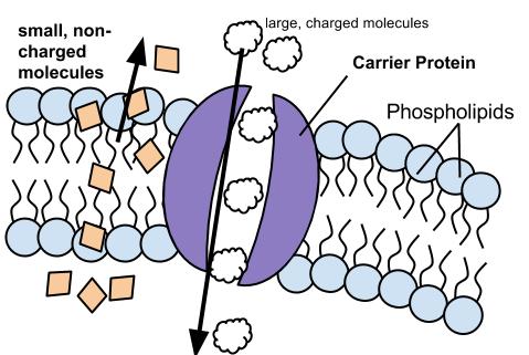
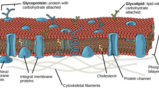
Cell membrane, thin membrane that surrounds every living cell. The cell membrane functions as a barrier, keeping cell constituents in and unwanted ...
Ch 5 Membrane Structure And Function
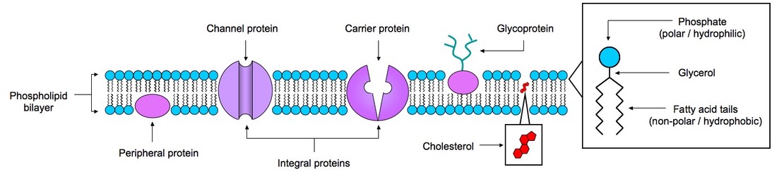
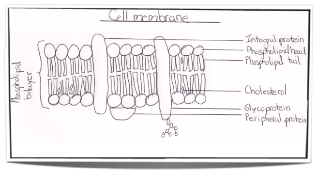
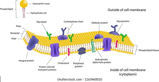
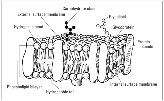
When drawing a diagram of a ... Our current model of the cell membrane is called the Singer-Nicholson fluid mosaic model ... Lines touch the labeled.31 pages

Cell Membrane Structure In Detail
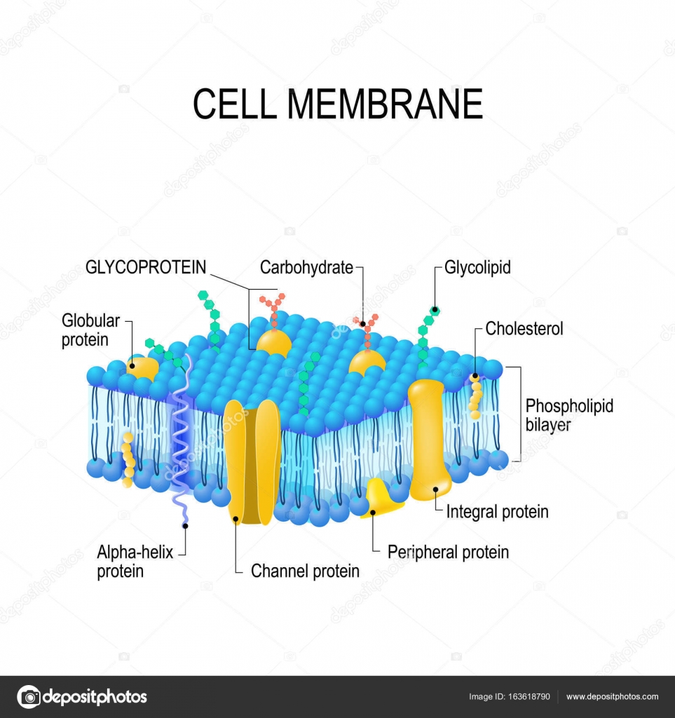
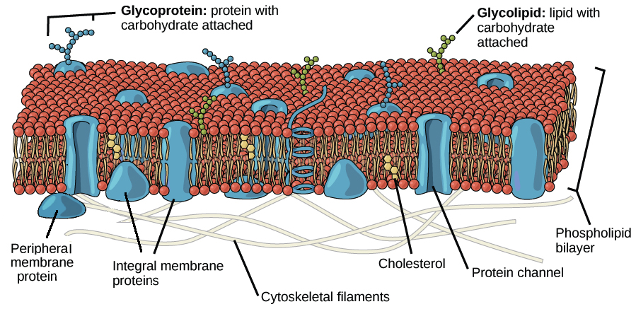
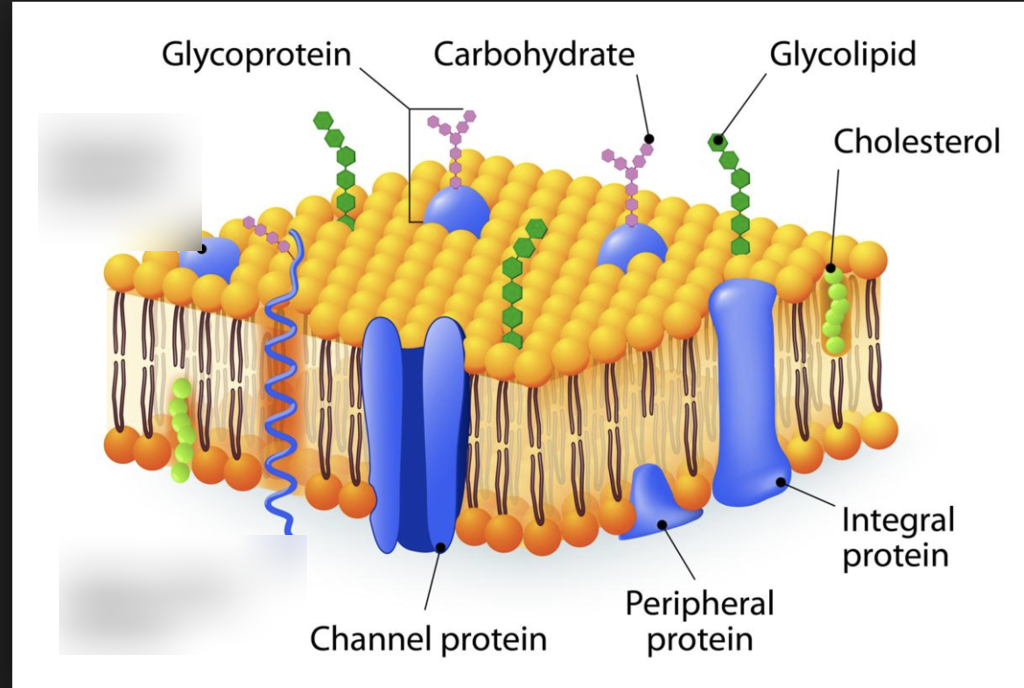
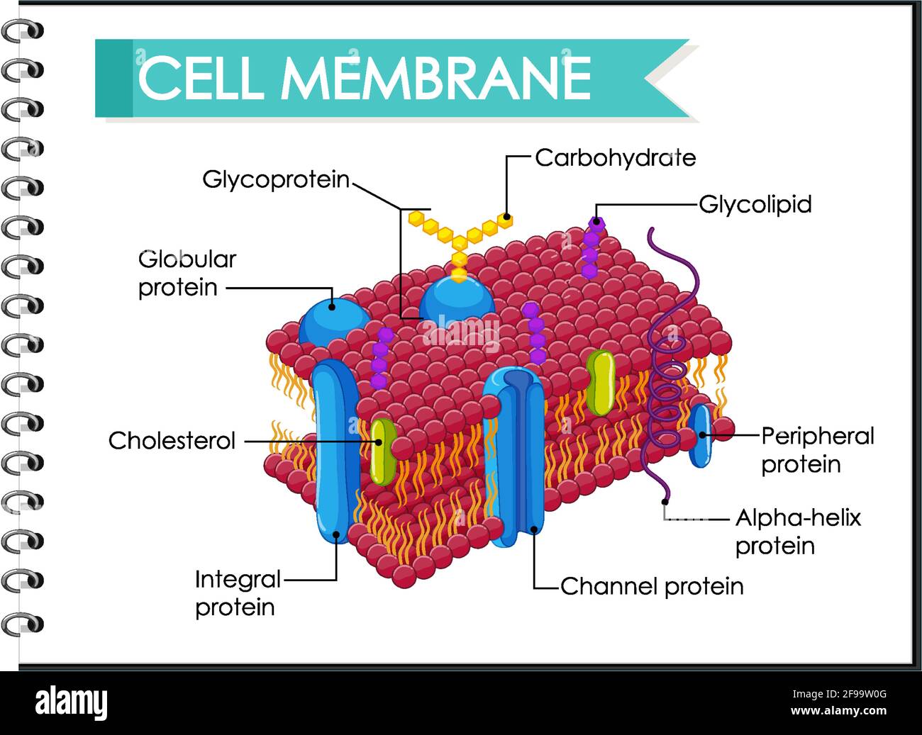
Aug 14, 2020 — The fluid mosaic model describes the plasma membrane structure as a mosaic of phospholipids, cholesterol, proteins, and carbohydrates.
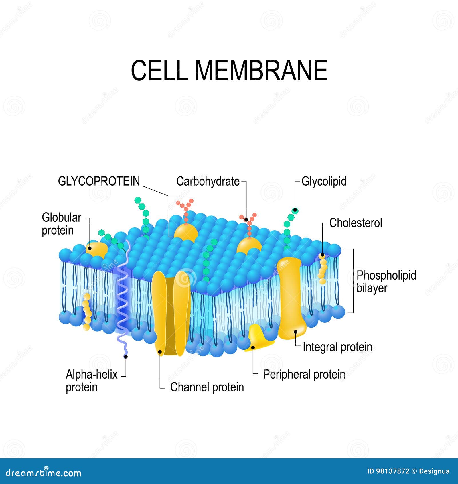
Cell Membrane Vector Stock Vector Image By C Edesignua 163618790
In this study, we synthesized a novel fluorescein isothiocyanate (FITC)-labeled prostate-specific membrane antigen (PSMA) ligand (PSMA-FITC) via the Fmoc solid-phase synthesis method, and the application value of PSMA-FITC in targeted fluorescence imaging of PSMA-positive prostate cancer was evaluated. The PSMA ligand developed based on the Glu-urea-Lys structure was linked to FITC by ...
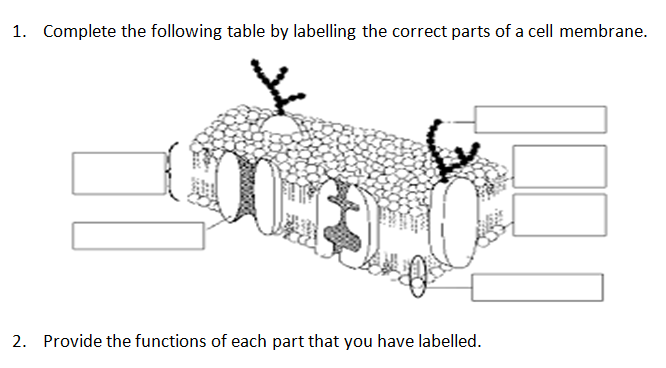
Answered 1 Complete The Following Table By Bartleby
Enhancing Cell Structure. Since human cell membranes are made with cholesterol, it's no surprise that cholesterol is needed for cell maintenance and creation. Specifically, cholesterol allows the cell membrane to stay flexible and allow lipids to pass through. Without cholesterol, cell walls are not properly shaped, which can cause problems ...
2 4 Membranes Bioninja
The oropharynx forms part of the pharynx, being the continuation of the oral cavity and nasopharynx superiorly, and the larynx and hypopharynx inferiorly. It also forms part of the upper respiratory tract and the gastrointestinal tract. Gross an...

2 3 Cell Structure And Function Cells The Basic Units Of Life Siyavula
Cell Structure Human Cell Diagram Cell Diagram Animal Cell Drawing Membranes And Membrane Lipids Cell Diagram Plant Cell Diagram Plant And Animal Cells Cotton Fiber Under A Microscope Schematic Drawing Fabric Textiles Cotton Https Www Researchgate Net Profile Parashuram Bannigidad Publication 260919435 Figure Fig1 As 3924188526 Bacterial Cell Structure Cell Structure Cell Diagram Cell Membrane
Ib Biology Notes 2 4 Membranes
The tympanic membrane should appear as an intact, ovoid, semitransparent, pearly gray membrane at the end of the canal. The lower four fifths of the tympanic membrane is called the pars tensa the upper fifth, the pars flaccida. The handle of the malleus should be seen near the center of the pars tensa.

Cell Membrane Wikipedia
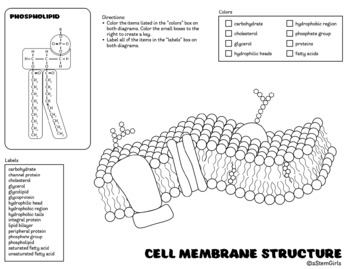
Finally, have students complete the Cell Membrane Color Sheet and Build a Cell Membrane activity to solidify their understanding of the cell's anatomy and functions. Cell Membrane Concepts Diffusion is a passive transport method of movement of molecules from higher concentration to lower concentration.

The Cell The Histology Guide
Jul 13, 2020 - This Pin was discovered by Michelle Figueroa. Discover (and save!) your own Pins on Pinterest.

Labeled Diagram Of Plasma Membrane Best Of Diagram With Plasma Membrane Stock Vector Illustration Plasma Membrane Cell Membrane Cell Membrane Structure
The lipid bilayer envelope, membrane proteins, and nucleocapsid protect the virus when it is outside the host cell. The viral envelope is made up of a lipid bilayer in which the membrane (M), envelope (E) and spike (S) structural proteins are anchored. The molar ratio of E:S:M in the lipid bilayer is approximately 1:20:300.
Label The Parts Of A Cell Membrane With Th Clutch Prep
Spindle fibers are structures that form during cell division. Explore the definition, location, and purpose of spindle fibers, and study an overview of cell division and spindle fiber formation.

35 Label The Plasma Membrane Labels Database 2020
An otoscope consists of a head and a handle and is used to examine the external auditory canal (EAC), the tympanic membrane, and the middle ear. A magnifying lens enhances the clinician's view ...
Structure Of The Plasma Membrane Article Khan Academy
29 Phospholipid Bilayer Diagram Label - Wiring Diagram List Molecular Graphic Of Membrane Phospholipid Bilayer Photograph by National Institutes Of Health ... Cell membrane lipid bilayer , artwork - Stock Image - F007/1479 - Science Photo Library
Cell Membrane Structure And Functions Diagram Quizlet
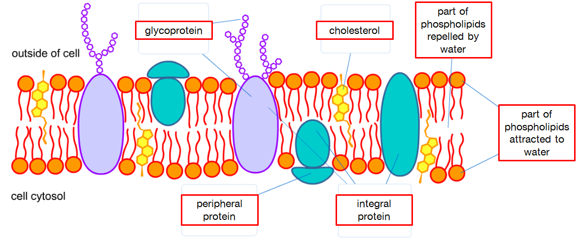
Understand the fluid mosaic model of membranes; Describe the functions of phospholipids, proteins, and carbohydrates in membranes. A cell's plasma membrane ...

Biology Quiz Structure And Function Of Cell Membrane Proprofs Quiz
Sodium is a chemical element with the symbol Na (from Latin natrium) and atomic number 11. It is a soft, silvery-white, highly reactive metal.Sodium is an alkali metal, being in group 1 of the periodic table. Its only stable isotope is 23 Na. The free metal does not occur in nature, and must be prepared from compounds.
Structure Of The Plasma Membrane Article Khan Academy
Lesson Summary. Keratin is an important protein in the epidermis. Keratin has two main functions: to adhere cells to each other and to form a protective layer on the outside of the skin. In ...

Membranes
(BOSTON) — The inherited progressive disorder cystic fibrosis (CF) causes severe damage to the lungs, and other tissues in the body by affecting the cells that produce mucus, sweat, and ...

Quia Ap Chapter 6 Cells Detailed
The respiratory system of the pig commences at the nostrils which lead into two nasal passages. These contain the dorsal and ventral turbinate bones. (Fig.1-8). The ventral turbinates consist of four thin main bones, two on each side separated by a cartilaginous septum. You can imagine these as four hair curlers placed inside the nose.
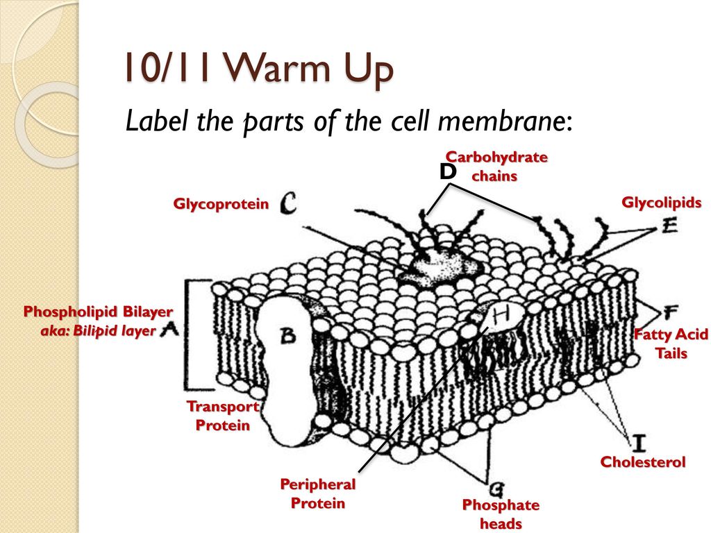
10 11 Warm Up Label The Parts Of The Cell Membrane D Carbohydrate Ppt Download
:max_bytes(150000):strip_icc()/plasma_membrane-58a617c53df78c345b5efb37.jpg)
Cell Membrane Function And Structure
Cell Membrane Detailed Diagram Labeled

2 4 1 Draw And Label A Diagram To Show The Structure Of Membranes Flashcards Quizlet
Human Cell Membrane Structure Illustration Stock Vector Image Art Alamy

Anatomy And Physiology Of Animals The Cell Wikibooks Open Books For An Open World
Cell Membrane Images Stock Photos Vectors Shutterstock
Cell Plasma Membrane Structure Color Label Perfect For Interactive Notebooks

Cell Membrane Definition Structure Function And Biology
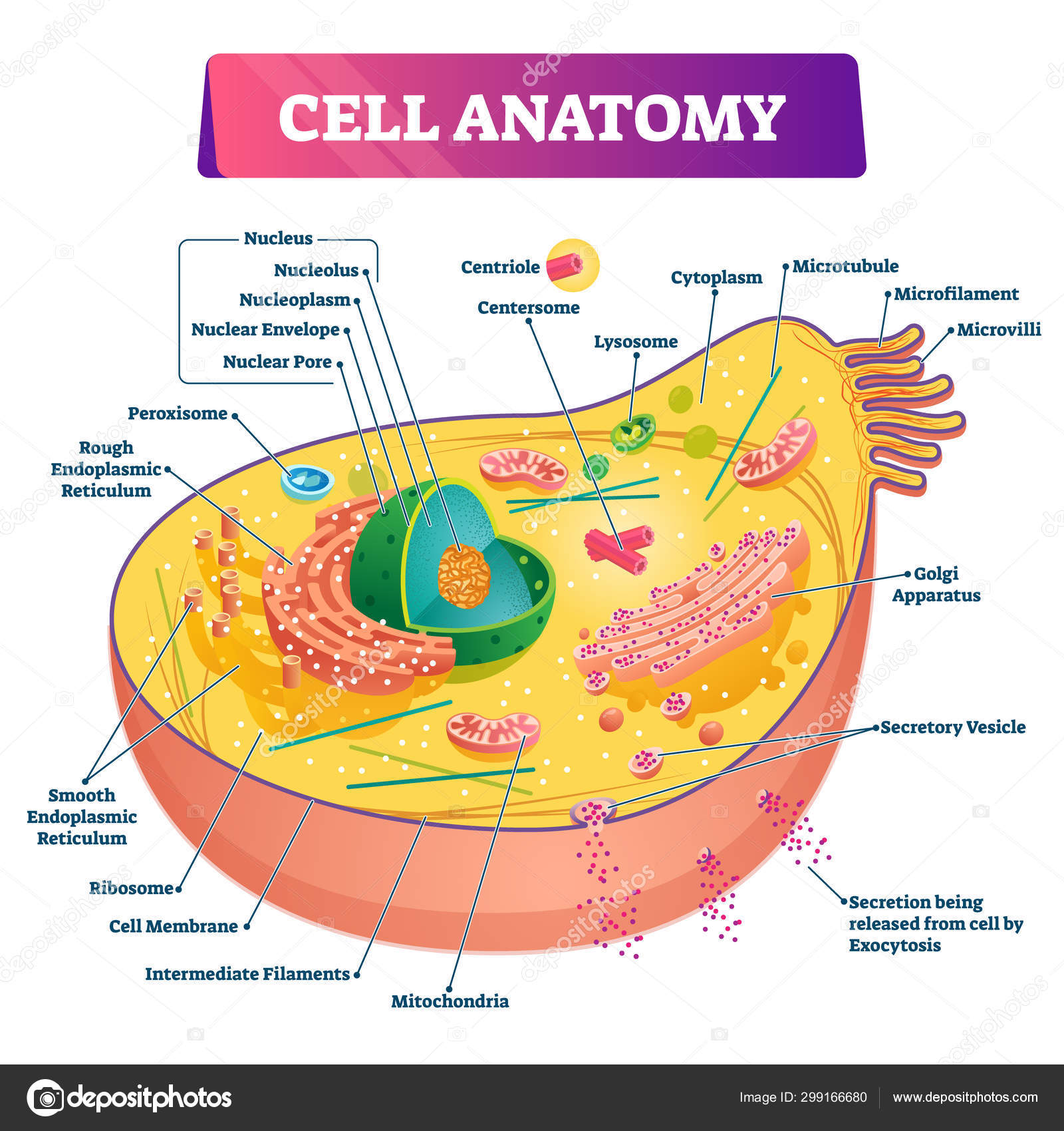
Cell Anatomy Vector Illustration Labeled Educational Structure Diagram Stock Vector Image By C Vectormine 299166680
Cell Membrane Stock Illustrations 4 738 Cell Membrane Stock Illustrations Vectors Clipart Dreamstime

3 1 The Cell Membrane Anatomy Physiology
1

The Cell Membrane Anatomy And Physiology I
The Fluid Mosaic Model Of The Cell Plasma Membrane Dummies

Cell Membrane Structure Diagram Quizlet
Cell Membrane Structure Practice Rachel Sanders Library Formative

3 1 The Cell Membrane Anatomy Physiology

Chapter4

Cell Membrane Pink Membrane Vector Royalty Free Cliparts Vectors And Stock Illustration Image 66990039
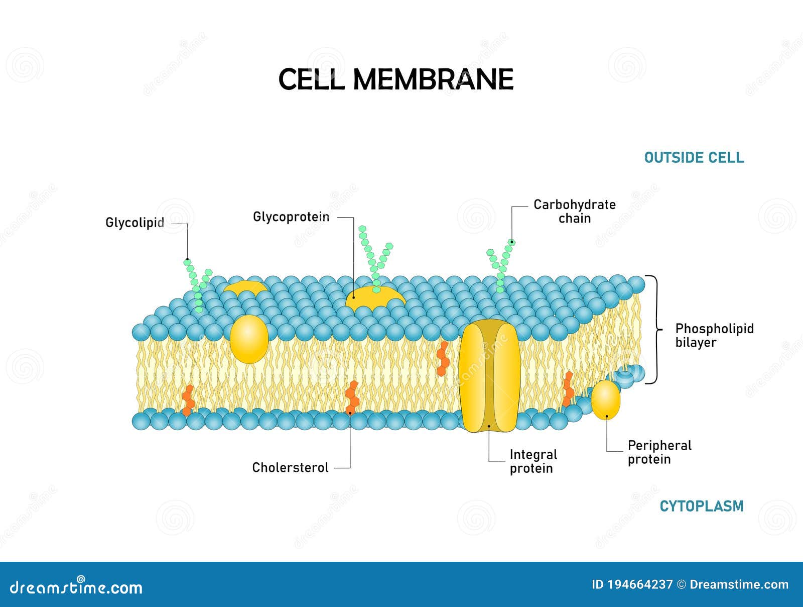
Diagram Of Cell Membrane Phospholipid Bilayers Structure Stock Vector Illustration Of Diffusion Biology 194664237
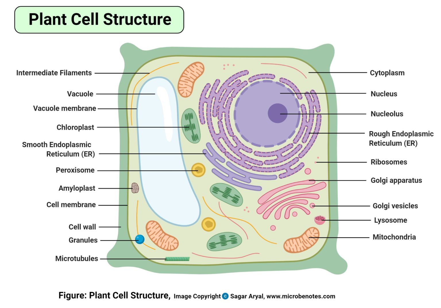
Plant Cell Definition Labeled Diagram Structure Parts Organelles


Comments
Post a Comment