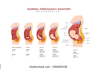
38 pregnant anatomy diagram
Anatomy of pregnancy and birth. 4-minute read. Listen. From the moment you find out that you (or your partner) are pregnant you may be filled with a rush of emotions. In addition, there is a range of physical changes that many pregnant women go through. Learn about the anatomical changes you can expect throughout conception, pregnancy and ... Anatomy of abdomen with twins. pregnancy type infographic elements in flat design. Monochorionic monoamniotic twins medical diagram isolated on white background. Fertilization diagram with human sperm, human egg and zygote, vector illustration.
Birth Anatomy - A Guide to Mother's Birth Anatomy - Spinning Babies. Mother's Birth-Related Anatomy. A woman's birthing anatomy includes soft tissues and hard bones. The bones. Our bones are held together by flexible tendons. In pregnancy, these joints become even more mobile. Waddling is an example of what happens when these joints get softer.

Pregnant anatomy diagram
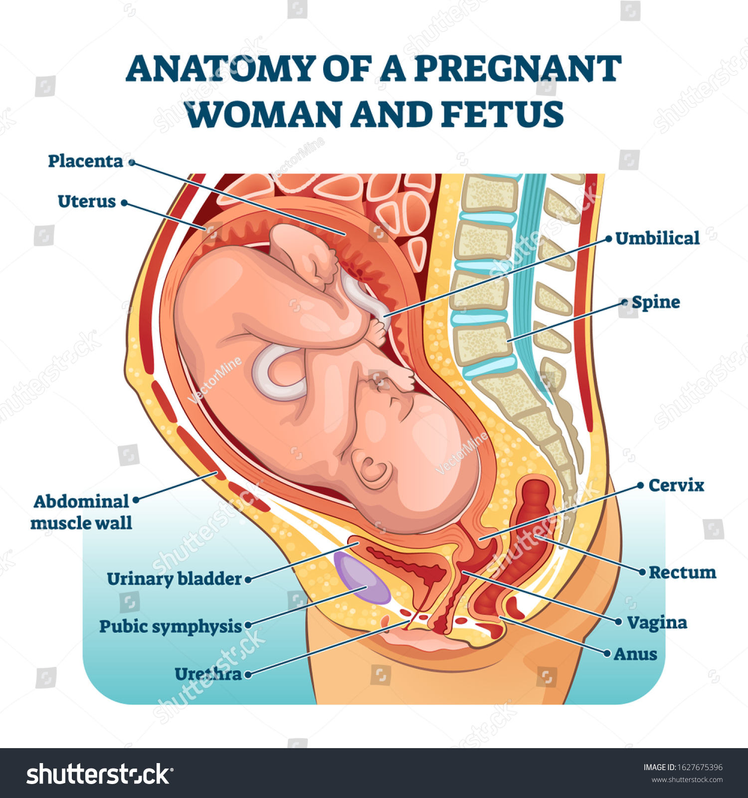
Let's begin by going over the reproductive anatomy of dogs and pointing out some unique features. Starting with the male. One unique feature of male dogs as compared to many other mammals is the presence of only one accessory sex gland. On this diagram, locate the prostate. The prostate gland is the only accessory sex gland of male dogs. First pregnancy, 32F, no issues during my pregnancy so far aside from a very short luteal phase which required for me to supplement with progesterone until week 12 to fall and stay pregnant . Also hashimoto thyroiditis, well controlled for years via meds I had a private anatomy scan at just over 19w and all was good. Then today at just over 23w I had my NHS scan at the hospital (I live in the UK) and they said there is probably a small cyst 'posterior to the cerellar vermis', in the cisterna ma... Description: Anatomy of a pregnant woman and fetus labeled diagram, vector illustration medical scheme. Family planning. Inner organs location in pregnancy. Abdominal muscle wall, bladder, vagina, cervix and other
Pregnant anatomy diagram. cross section of the mothers anatomy at 9 months showing the baby in uteruo loa ready to be delivered - diagram of pregnancy stock illustrations pregnant businesswoman presenting new plans - diagram of pregnancy stock pictures, royalty-free photos & images Browse 230 uterus diagram stock photos and images available or start a new search to explore more stock photos and images. An anatomical diagram depicts the method of extracting a fetus by reaching into the uterus and adjusting the baby's position. Diagram Of A Human Uterus During The Seventh Week Of Pregnancy. I just found out I was pregnant and am terrified of a VBAC but also a csection. My first csection the spinal block was administered and within what seemed like seconds I went numb from the neck down and felt like just a head. I went into full blown shock as I couldn’t feel myself breathing. And was unable to breathe Will this happen again? Was this a placement issue? Or was this anatomy? Find Pregnant Fetus Anatomy Diagram Illustration stock images in HD and millions of other royalty-free stock photos, illustrations and vectors in the Shutterstock collection. Thousands of new, high-quality pictures added every day.
Anatomy: Fetus in Utero. Amniotic sac. A thin-walled sac that surrounds the fetus during pregnancy. The sac is filled with liquid made by the fetus (amniotic fluid) and the membrane that covers the fetal side of the placenta (amnion). This protects the fetus from injury. it also helps to regulate the temperature of the fetus. Posted on September 26, 2014 by admin. week pregnancy pic. Week Pregnancy Pic Diagram - Week Pregnancy Pic Chart - Human anatomy diagrams and charts explained. This diagram depicts Week Pregnancy Pic with parts and labels. Mpreg Anatomy. Published: Aug 19, 2012. By. althea9. 199 Favourites. 81 Comments. 16K Views. well, it was a long time i wanted do this anatomical table, and so, this is my personal explanation of male pregnancy, from the conception at the birth! if you wont, say me your ideas about it! [https://twitter.com/GLITTER\_TENDON/status/1481711404457594885](https://twitter.com/GLITTER_TENDON/status/1481711404457594885) for the idea!! https://preview.redd.it/cnleiqjo3qc81.jpg?width=3360&format=pjpg&auto=webp&s=435d0910a72f425192a24c2eb23b0533dcf800d2
diagrams giving them all the correct answers. Use the anatomy posters to label all the parts of the male and female reproductive systems. While the students follow along and continue to label their sheets, explain the parts of the male anatomy. Remind students that the brain is the most important part of both the male and the Pregnant fetus anatomy diagram. Pregnant with fetus. Pregnant with fetus. Pregnant anatomy with fetus. Pregnant anatomy with fetus. Pregnant anatomy and the fetus. More stock photos from Blueringmedia's portfolio. A Baby in Mother Womb. Little baby curl up. An Education Poster of Fetal Development. ANATOMICAL CHANGES IN PREG uUterus u4 weeks:Enlarged and globular, increasing in size by about 1 cm per week u6 weeks: Uterus softens u12 weeks: uterus is sufficiently large to palpate abdominally just above the pubic symphysis u20 weeks: top of the uterus is at the umbilicus u> 20 weeks: fundal height can be used as measurement of GA by measuring distance from pubic symphisisto top most ... iStock Fetus In Utero Pregnancy Women Diagrams Pregnant Female Anatomy Stock Illustration - Download Image Now Download this Fetus In Utero Pregnancy Women Diagrams Pregnant Female Anatomy vector illustration now. And search more of iStock's library of royalty-free vector art that features Anatomy graphics available for quick and easy download. Product #: gm1156246298 $ 12.00 iStock In stock
Female pregnant anatomy Images, Stock Photos & Vectors ...
Female anatomy refers to the internal and external structures of the reproductive and urinary systems. Reproductive anatomy aids with sexual pleasure, getting pregnant, and breastfeeding a baby. The urinary system helps rid the body of toxins through urination (peeing).

Female Reproductive Anatomy, Pregnancy Birth Poster: Amazon ...
What does it mean when your baby has a too much fluid in there brain Does this cause any health problems to me or the baby? Has anyone else experience this if so what did you do? Has anyone experience a ventriculomegaly pregnancy?

Clip Art Vector - Normal pregnant female anatomy. Stock EPS ...
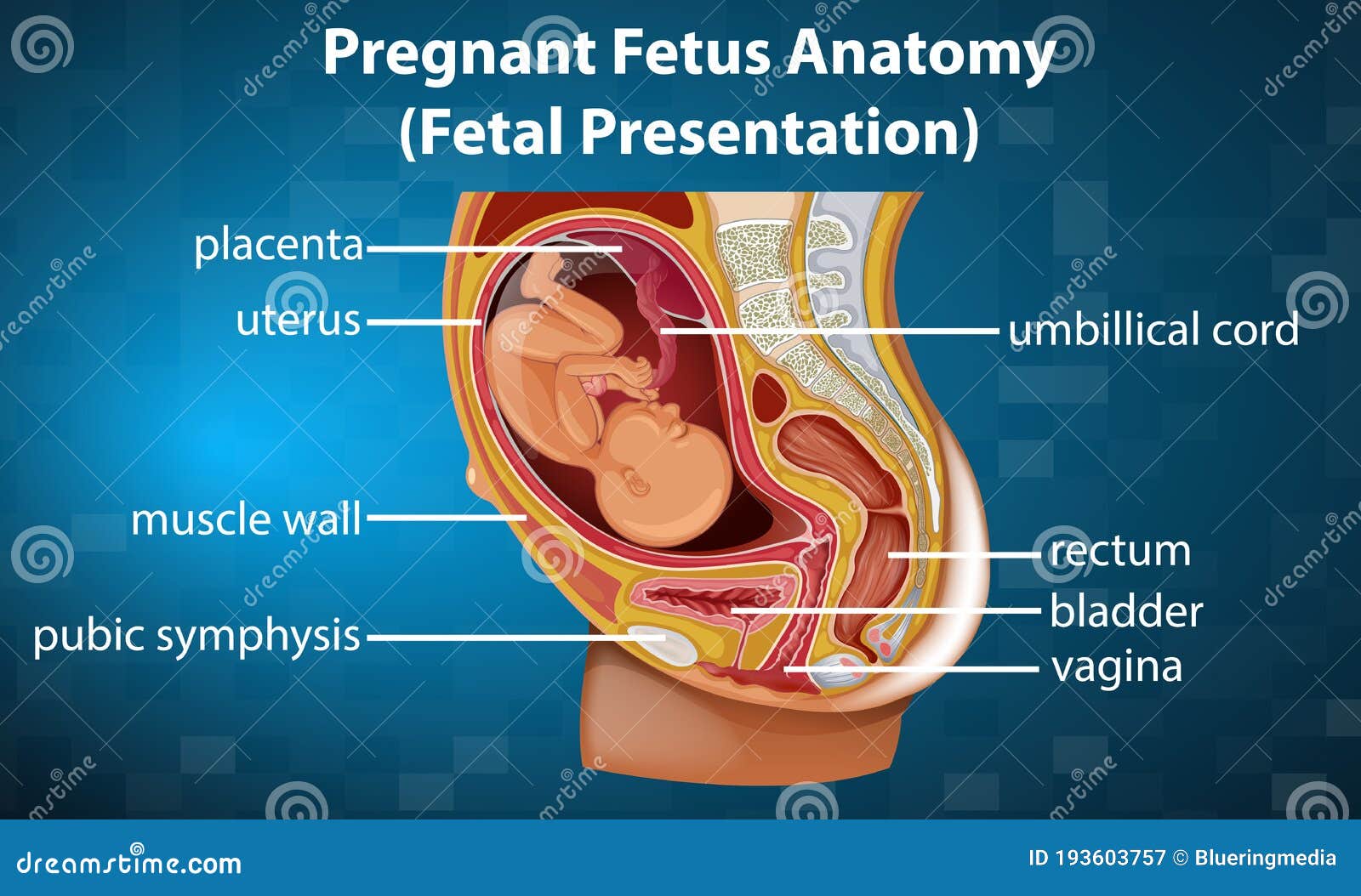
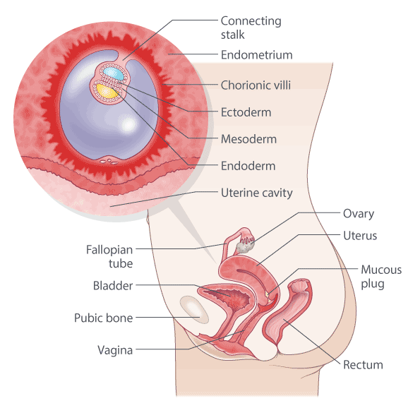
The diagram shows part of the internal anatomy of a pregnancy focusing on the embryo, extraembryonic membranes, and other essential physiology needed for fetal development.

Female Reproductive Anatomy, Pregnancy Birth Anatomy Chart Uterus Model
pregnant female anatomy diagram. A 29-year-old female asked: if on medical aid is an anatomy scan a must to do during pregnancy? Dr. Brad Douglas answered. 24 years experience Obstetrics and Gynecology. Recommended: Yes it is recommended around 20 weeks of pregnancy . Send thanks to the doctor.
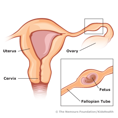
Ectopic Pregnancy (for Parents) - Nemours KidsHealth
The vagina is an elastic, muscular canal with a soft, flexible lining that provides lubrication and sensation. The vagina connects the uterus to the outside world. The vulva and labia form the ...
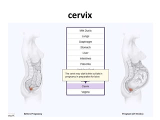
Anatomical changes in Pregnancy
Download this Free Vector about Pregnant fetus anatomy diagram, and discover more than 20 Million Professional Graphic Resources on Freepik. #freepik #vector #education #medical #science

Pregnant fetus anatomy diagram 1591218 Vector Art at Vecteezy
Download this Free Vector about Pregnant fetus anatomy diagram, and discover more than 19 Million Professional Graphic Resources on Freepik. #freepik #Vector #Baby #Cartoon #Kids

Pregnancy Week 23 Pregnant Woman And Infant Anatomy
More similar stock illustrations. Visible uterus and fetus week 40. Pregnant woman. Visible uterus and fetus week 36. Illustration of a baby in the womb. Pregnant with fetus. silhouette of pregnant woman isolated on white background. Pregnant fetus anatomy diagram. Pregnant fetus anatomy diagram.
Anatomical changes during pregnancy | Kenhub
2 Reproductive Anatomy of the Cow/Heifer An important feature of the cow/heifer reproductive tract is its location. Manipulation of the reproductive tract for various management practices, such as rectal palpation for pregnancy diagnosis or artifcial insemination, is

Pregnant Woman Body High-Res Vector Graphic - Getty Images
How this works. Pregnancy is a time of great physical and emotional change for women. Everything from belly size to heartbeat speed will change over the 9 months leading up to childbirth. Partly ...

1 Weeks Pregnancy Diagram
​ https://preview.redd.it/m7tdq980bve71.jpg?width=3470&format=pjpg&auto=webp&s=032178c725512ea76cd7d69232fe7118b6eeb734

Pregnant women anatomical models. Pregnant woman models at 3 ...
Your Body at 6-7 Weeks of Pregnancy. When you are between 6 and 7 weeks pregnant, you may be experiencing the early signs of pregnancy: your period has stopped and you may have nausea, breast tenderness and swelling, frequent urination and fatigue. At this point, your uterus has begun to grow and become more egg-shaped.
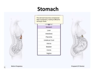
Anatomical changes in Pregnancy
Pregnancy women diagrams Pregnant female anatomy. Female reproductive system, image diagram uterus diagram stock pictures, royalty-free photos & images. Female reproductive system, image diagram. female reproductive system Vector female reproductive system, flat design uterus diagram stock illustrations.

Pregnancy Illustrations | Visualisations of the Human ...
a woman who is 30 weeks pregnant with chalkboard & information about pregnancy - female anatomy diagram stock pictures, royalty-free photos & images. medical illustration of man and woman - female anatomy diagram stock illustrations.

Female pregnant anatomy Images, Stock Photos & Vectors ...
The pelvis is located in the middle of the human body, below the abdomen and above the thighs. It comprises the bony pelvis — which includes the hip bones —, pelvic cavity, pelvic floor, and perineum — the area of skin between the opening of the vagina and the anus. The bones around the pelvis are attached by several ligaments, comprised ...

Pregnancy Week-by-Week Early and Later Signs & Symptoms
The cervix is a fibromuscular organ that links the uterine cavity to the vagina. Although it is described as being cylindrical in shape, the anterior and posterior walls are more often ordinarily apposed. The cervix is approximately 4 cm in length and 3 cm in diameter. The cervix of a parous woman is considerably larger than that of a nulliparous woman, and the cervix of a woman of ...
Pregnant Fetus Anatomy Diagram Stock Vector - Illustration of ...
Description: Anatomy of a pregnant woman and fetus labeled diagram, vector illustration medical scheme. Family planning. Inner organs location in pregnancy. Abdominal muscle wall, bladder, vagina, cervix and other

34 Weeks Pregnancy Diagram
First pregnancy, 32F, no issues during my pregnancy so far aside from a very short luteal phase which required for me to supplement with progesterone until week 12 to fall and stay pregnant . Also hashimoto thyroiditis, well controlled for years via meds I had a private anatomy scan at just over 19w and all was good. Then today at just over 23w I had my NHS scan at the hospital (I live in the UK) and they said there is probably a small cyst 'posterior to the cerellar vermis', in the cisterna ma...
Female pregnant anatomy Images, Stock Photos & Vectors ...
Let's begin by going over the reproductive anatomy of dogs and pointing out some unique features. Starting with the male. One unique feature of male dogs as compared to many other mammals is the presence of only one accessory sex gland. On this diagram, locate the prostate. The prostate gland is the only accessory sex gland of male dogs.

Pregnant fetus anatomy diagram - Stock Illustration [68957341 ...

Pregnant anatomy belly Images, Stock Photos & Vectors ...

Pregnant anatomy belly Images, Stock Photos & Vectors ...

Pregnancy Illustrations | Visualisations of the Human ...
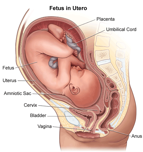
Anatomy: Fetus in Utero

Ectopic pregnancy: Signs, treatments, causes, risk factors ...

Pregnant Womb Diagram Stock Illustrations – 93 Pregnant Womb ...

13 Weeks Pregnant Anatomy Sectional View

13 Weeks Pregnancy Diagram
Anatomy Pregnant Woman Fetus Labeled Diagram Stock Vector ...
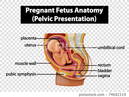
Pregnant Fetus Anatomy (Pelvic Presentation) - Stock ...

Anatomy of Abdomen with Twins. Pregnancy Women Diagrams with ...

Pin on Pregnancy
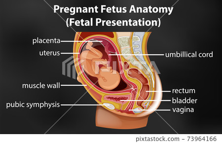
Pregnant fetus anatomy diagram - Stock Illustration [73964166 ...

Digital Illustration Of Pregnant Anatomy With Fetus In Colour ...
4 weeks pregnant | Raising Children Network

Female pregnant anatomy Images, Stock Photos & Vectors ...
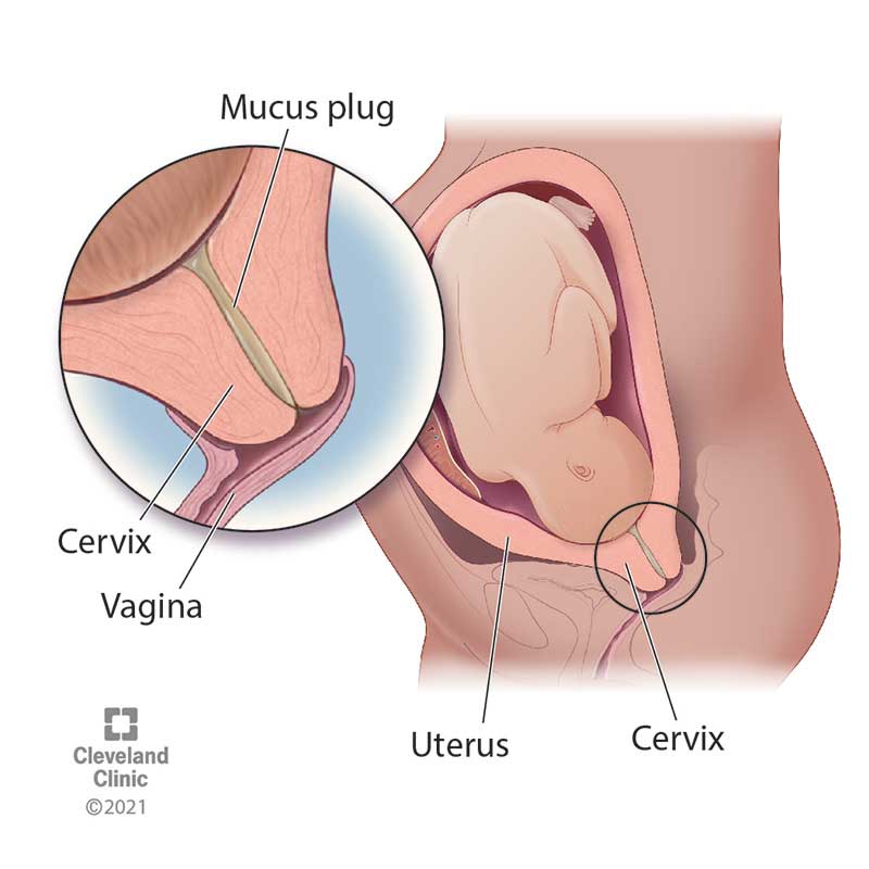
Mucus Plug: What It Is, Looks Like & Means

Pregnant 23 Weeks Diagram


Comments
Post a Comment